
You have no items in your shopping cart.
Cell Biology & Pathology
Epithelial Cell Markers
Essential targets for identifying epithelial lineages, studying the epithelial-mesenchymal transition (EMT), and isolating circulating tumor cells.
1. Biological Overview
Epithelial tissues cover external body surfaces and line many internal organs, ducts, and body cavities; blood vessels are lined by specialized endothelial cells. They serve critical functions, including providing a physical protective barrier, facilitating secretion (glandular epithelium), and managing absorption (intestinal epithelium). Because epithelial cells are tightly packed and physically connected via structural junctions, understanding their unique molecular signatures is critical for histology and cell biology.
Epithelial cell markers are proteins preferentially or characteristically expressed by epithelial lineages and are used collectively to identify epithelial differentiation. These markers are indispensable in research and diagnostics, allowing scientists to identify the tissue of origin for metastatic carcinomas, evaluate structural integrity, and study cellular plasticity during embryogenesis and cancer progression.
2. Key Epithelial Markers
Researchers utilize a variety of structural and functional proteins to classify epithelial cells depending on the specific tissue type and experimental context. Explore our extensive portfolio by clicking on the marker categories below.
Cytokeratins (CKs)
Cytokeratins form the intermediate filament network within epithelial cells. Human keratins comprise a large family of proteins whose expression patterns are strongly dependent on epithelial type, differentiation state, and tissue. For example, CK8 and CK18 are characteristic of simple single-layered epithelia, whereas broad-spectrum "Pan-Cytokeratin" antibodies (like the AE1/AE3 clone) are routinely used to confirm epithelial origin in unknown neoplasms.
E-Cadherin (CD324)
E-Cadherin is a calcium-dependent cell adhesion molecule essential for forming adherens junctions. It maintains the physical integrity of epithelial sheets and acts as a tumor suppressor. Reduced or dysfunctional E-cadherin is frequently associated with tumor progression, invasion, and metastasis, although E-cadherin loss is not universally required for these processes.
EpCAM (CD326)
The Epithelial Cell Adhesion Molecule (EpCAM) is a pan-epithelial marker highly expressed in carcinomas but virtually absent in mesenchymal cells. EpCAM is one of the most widely used epithelial targets for CTC enrichment, but EpCAM-dependent methods can miss CTCs with low or absent EpCAM expression, particularly cells undergoing EMT or displaying mesenchymal/hybrid phenotypes.
Tight Junction Proteins
Proteins such as Claudins, Occludin, and ZO-1 (Zonula Occludens-1) govern the paracellular permeability of epithelial layers. They are critical markers when evaluating barrier function in in vitro models (e.g., Caco-2 cell permeability assays) and studying pathogen entry.
3. Epithelial-Mesenchymal Transition (EMT) and Pathology
The Epithelial-Mesenchymal Transition (EMT) is a fundamental biological process wherein polarized, immotile epithelial cells undergo biochemical changes to adopt a mesenchymal cell phenotype, characterized by enhanced migratory capacity and invasiveness.
During EMT, cells experience the "Cadherin Switch"—a profound downregulation of epithelial markers (E-Cadherin, Cytokeratins, ZO-1) coupled with the upregulation of mesenchymal markers (N-Cadherin, Vimentin, Fibronectin, and transcription factors like Snail and Slug).
Pathological activation of EMT-related programs can contribute to fibrosis, invasion, and metastasis. Monitoring coordinated changes in multiple epithelial and mesenchymal markers can help characterize EMT-related phenotypes, provided that marker selection and interpretation are validated for the experimental system.
4. Featured Research Reagents
Biorbyt offers a comprehensive portfolio of highly validated antibodies to interrogate epithelial biology, structural integrity, and EMT pathways.
| Image | Product Details | Application & Reactivity |
|---|---|---|
|
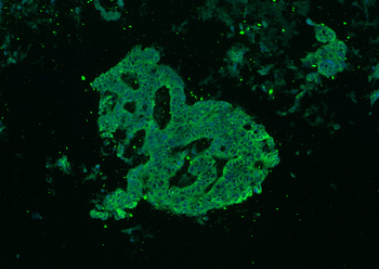
Pan-Cytokeratin Rabbit Polyclonal
SKU: orb10399
Broad-spectrum marker cocktail is widely used to identify carcinomas and confirm epithelial lineage. |
Applications: ELISA, IHC, WB, IF
Reactivity: Human, Mouse, Rat, Bovine, Dog |
|
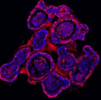
Anti-E-Cadherin Antibody
SKU: orb308856
Critical for cell-cell adhesion. Monitored closely to detect the onset of the Epithelial-Mesenchymal Transition. |
Applications: IHC, WB, ELISA
Reactivity: Human, Mouse, Rat |
|
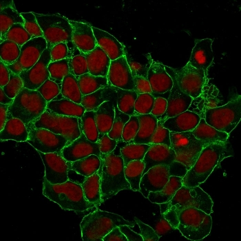
EpCAM Antibody
SKU: orb606357
Pan-epithelial carcinoma marker utilized for diagnosing metastasis and capturing Circulating Tumor Cells (CTCs). |
Applications: IHC, WB, Flow Cytometry
Reactivity: Human, Mouse, Rat |
|
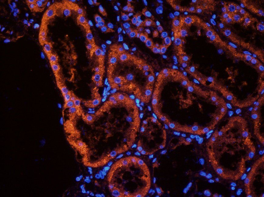
MUC1 Antibody
SKU: orb315622
A heavily glycosylated transmembrane protein expressed on the apical surface. Overexpressed in adenocarcinomas. |
Applications: IF/ICC, IHC, WB
Reactivity: Human, Mouse, Rat |
|
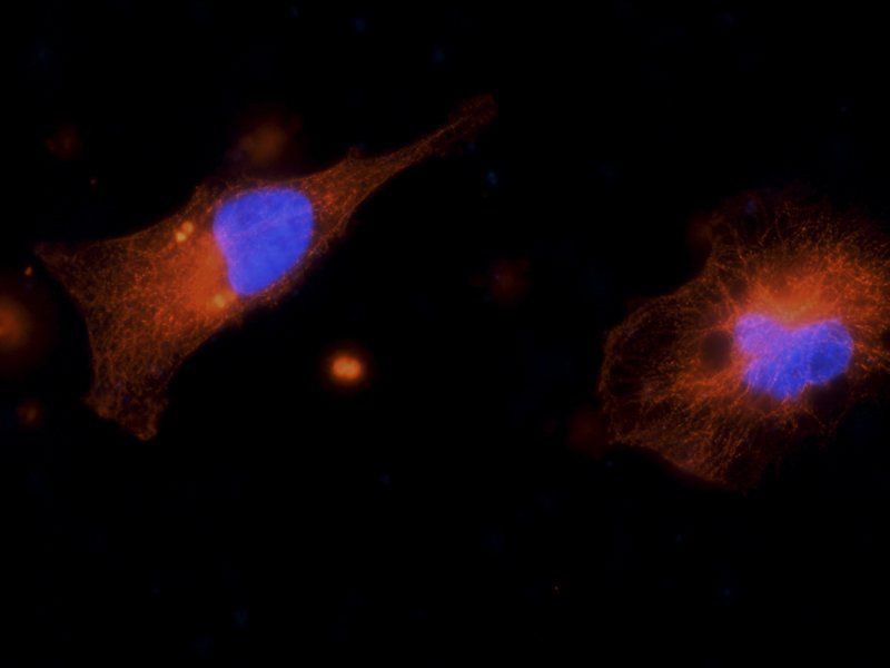
ZO-1 Antibody
SKU: orb313868
Peripheral membrane protein that anchors tight junction strands. Essential for assessing in vitro barrier integrity. |
Applications: IF, IHC, WB
Reactivity: Human, Mouse, Rat |
|
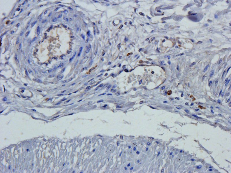
Claudin-1 Antibody
SKU: orb10447
Integral component of epithelial tight junctions regulating paracellular transport and barrier defense. |
Applications: IHC, WB, ELISA
Reactivity: Human, Mouse, Rat |
5. Custom Antibody & Protein Services
Studying a novel epithelial splice variant, requiring unique species reactivity, or navigating complex transmembrane targets? Biorbyt provides tailored contract research services to overcome experimental bottlenecks.
- Custom Nanobody Development Service: Biorbyt provides custom single-domain antibody (VHH) development from immunized camelids (alpacas and llamas). Using a proprietary Single B platform, rapid library generation, and high-throughput screening, validated nanobody hits can be identified within 2 months.
- Functional Membrane Protein Expression: Overcome structural bottlenecks with high-yield native-conformation preparation of complex transmembrane targets like GPCRs, Ion Channels, Transporters, and Extracellular Domains (ECDs). Biorbyt utilizes optimized platforms such as MSP Nanodiscs, PeptiNanodiscs, Membrane Nanoparticles (MNP), and Virus-Like Particles (VLP).
- Custom Peptide Synthesis & Recombinant Antibodies: Biorbyt also offers bespoke peptide synthesis (up to 100 amino acids) with diverse modifications (PEGylation, glycosylation, fluorescent labeling, D-amino acids) alongside completely sequence-defined custom recombinant antibody production.
Need custom solutions for your epithelial research?
Reach out to Biorbyt’s scientific support team to discuss custom synthesis, specialized nanobody development, or membrane protein expression to accelerate your project.
Contact Technical Support6. Frequently Asked Questions
What is the best pan-epithelial marker for identifying carcinomas?
Pan-Cytokeratin antibody cocktails (such as the AE1/AE3 clone blend) are widely used in diagnostic immunohistochemistry to identify cells of epithelial origin and classify carcinomas.
How do markers change during the Epithelial-Mesenchymal Transition (EMT)?
Cells undergoing EMT-related changes often reduce expression of epithelial markers and increase expression of selected mesenchymal markers, although partial/hybrid EMT states can retain both.The E-cadherin → N-cadherin shift is commonly called the "cadherin switch".
Why is EpCAM used for Circulating Tumor Cell (CTC) isolation?
EpCAM is universally expressed on the surface of normal epithelial cells and the vast majority of carcinomas, but is absent on blood cells (leukocytes). This distinct expression profile makes anti-EpCAM antibodies suitable for capturing and isolating rare CTCs from whole blood using microfluidic or magnetic sorting platforms.
What is the difference between adherens junctions and tight junctions?
Adherens junctions (characterized by E-cadherin) primarily provide strong mechanical attachments between adjacent epithelial cells. Tight junctions (characterized by Claudins, Occludin, and ZO-1) are located closer to the apical surface and function primarily to seal the intercellular space, regulating the passage of ions and molecules (barrier function).
7. References
- Thiery, J. P., Acloque, H., Huang, R. Y., & Nieto, M. A. (2009). Epithelial-mesenchymal transitions in development and disease. Cell, 139(6), 871-890.
- Moll, R., Divo, M., & Langbein, L. (2008). The human keratins: biology and pathology. Histochemistry and cell biology, 130(6), 705-733.
- Gires O, Pan M, Schinke H, Canis M, Baeuerle PA. Expression and function of epithelial cell adhesion molecule EpCAM: where are we after 40 years? Cancer Metastasis Reviews. 2020;39(3):969–987. DOI: 10.1007/s10555-020-09898-3.