You have no items in your shopping cart.
CD31 PECAM1 Rabbit Monoclonal Antibody
SKU: orb547772
Description
Images & Validation
−Item 1 of 2
| Tested Applications | FC, IHC, IP, WB |
|---|---|
| Reactivity | Human |
| Application Notes |
Key Properties
−| Antibody Type | Primary Antibody |
|---|---|
| Host | Rabbit |
| Clonality | Monoclonal |
| Isotype | Rabbit IgG |
| Clone No. | CEE-16 |
| Immunogen | A synthesized peptide derived from human CD31 |
| Molecular Weight | 120-130 kDa |
| Purification | Affinity-chromatography |
| Conjugation | Unconjugated |
Storage & Handling
−| Storage | Maintain refrigerated at 2-8°C for up to 2 weeks. For long term storage store at -20°C in small aliquots to prevent freeze-thaw cycles. |
|---|---|
| Form/Appearance | Liquid |
| Concentration | 0.5mg/ml |
| Disclaimer | For research use only |
Alternative Names
−Platelet endothelial cell adhesion molecule; PECAM-1; EndoCAM; GPIIA'; PECA1; CD31; PECAM1
Similar Products
−- Item 1 of 4
CD31 Recombinant Rabbit Monoclonal Antibody [orb1183945]
FC, ICC, IF, IHC-Fr, IHC-P, WB
Human
Rabbit
Recombinant
Unconjugated
25 μl, 100 μl, 50 μl - Item 1 of 2
- Item 1 of 1
Recombinant CD31 / PECAM-1 (Endothelial Cell Marker) Antibody [orb389189]
FC, IHC, WB
Human
Rabbit
Monoclonal
Unconjugated
100 μg (without BSA and Azide), 100 μg, 20 μg - Item 1 of 1
CD31 PECAM1 Rabbit Monoclonal Antibody [orb547773]
FC, IHC, WB
Human
Rabbit
Monoclonal
Unconjugated
100 μl
![Anti-CD31 [BAG-85D10]](/images/pub/media/catalog/product/NewWebsite/35/orb758909_1.png)
![Anti-CD31 [BAG-85D10]](/images/pub/media/catalog/product/NewWebsite/35/orb758909_2.png)


Quality Guarantee
Explore bioreagents carefree to elevate your research. All our products are rigorously tested for performance. If a product does not perform as described on its datasheet, our scientific support team will provide expert troubleshooting, a prompt replacement, or a refund. For full details, please see our Terms & Conditions and Buying Guide. Contact us at [email protected].

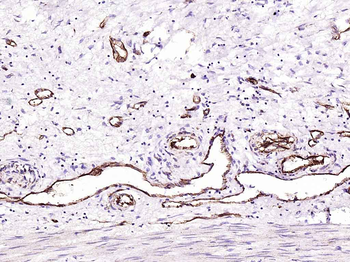
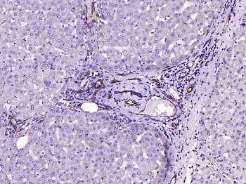
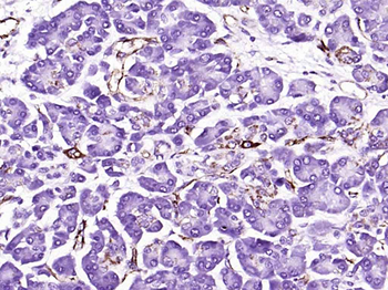
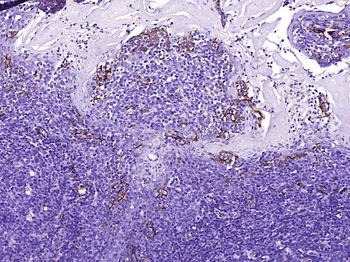
Immunohistochemical analysis of paraffin-embedded human kindey, using CD31 Antibody (orb547772) PECAM1 was detected in paraffin-embedded tissue section. Heat mediated antigen retrieval was performed in citrate buffer (pH6, epitope retrieval solution) for 20 mins. The tissue section was blocked with 10% goat serum. The tissue section was then incubated with 1ug/ml rabbit anti-PECAM1 Antibody (orb547772) overnight at 4°C. Biotinylated goat anti-rabbit IgG was used as secondary antibody and incubated for 30 minutes at 37°C. The tissue section was developed using Strepavidin-Biotin-Complex (SABC) (Catalog # orb90444) with DAB as the chromogen.

Western blot analysis of CD31 expression in THP-1 cell lysates (orb547772). Electrophoresis was performed on a 5-20% SDS-PAGE gel at 70V (Stacking gel)/90V (Resolving gel) for 2-3 hours. The sample well of each lane was loaded with 50ug of sample under reducing conditions. After Electrophoresis, proteins were transferred to a Nitrocellulose membrane at 150mA for 50-90 minutes. Blocked the membrane with 5% Non-fat Milk/TBS for 1.5 hour at RT. The membrane was incubated with rabbit anti-PECAM1 monoclonal antibody (Catalog # orb547772) overnight at 4°C, then washed with TBS-0.1%Tween 3 times with 5 minutes each and probed with a goat anti-rabbit IgG-HRP secondary antibody at a dilution of 1:10000 for 1.5 hour at RT. The signal is developed using an Enhanced Chemiluminescent detection (ECL) kit (Catalog # orb90503) with Tanon 5200 system. A specific band was detected for PECAM1.
Quick Database Links
UniProt
UniProt Details
− No UniProt data available
Documents Download
Datasheet
Product Information
Request a Document
Protocol Information
WB
Western Blot (IB, immunoblot)
IHC
Immunohistochemistry
FC
Flow Cytometry
IP
Immunoprecipitation
CD31 PECAM1 Rabbit Monoclonal Antibody (orb547772)

- 0.0
Based on 0 reviews
Participating in our Biorbyt product reviews program enables you to support fellow scientists by sharing your firsthand experience with our products.
Login to Submit a ReviewAvailable Sizes
Select a size below