You have no items in your shopping cart.
Ep-CAM / CD326 (Extracellular Domain) (Epithelial Marker) Antibody
SKU: orb1410486
Description
Images & Validation
−Item 1 of 10
| Tested Applications | ELISA, FC, IHC, WB |
|---|---|
| Dilution range | Flow Cytometry (0.5-1ug/million cells); Immunofluorescence (1-2ug/ml); ,Western Blotting (1-2ug/ml); ,Immunohistology (Formalin-fixed) (1-2ug/ml for 30 min at RT),(Staining of formalin-fixed tissues requires boiling tissue sections in 10mM Citrate Buffer, pH 6.0, for 10-20 min followed by cooling at RT for 20 minutes),Optimal dilution for a specific application should be determined. |
| Reactivity | Human |
| Application Notes |
Key Properties
−| Host | Mouse |
|---|---|
| Clonality | Monoclonal |
| Isotype | IgG1 |
| Clone No. | EGP40/1372 |
| Immunogen | Recombinant fragment from the extracellular domain of human EpCAM protein (around aa100-224) (exact sequence is proprietary) |
| Target | EPCAM |
| Source/Expression System | Bioreactor |
| Molecular Weight | 40-43kDa |
| Purification | Protein G |
| Conjugation | HRP |
Storage & Handling
−| Storage | Maintain refrigerated at 2-8°C for up to 2 weeks. For long term storage store at -20°C in small aliquots to prevent freeze-thaw cycles. |
|---|---|
| Form/Appearance | 200ug/ml of Ab purified from Bioreactor Concentrate by Protein A/G. Prepared in 10mM PBS with 0.05% rAlbumin & 0.05% azide. Also available WITHOUT rAlbumin & azide at 1.0mg/ml. |
| Buffer/Preservatives | Antibody purified from Bioreactor Concentrate by Protein A/G and conjugated to various reporter molecules. Prepared in 10mM PBS with 0.05% rAlbumin and 0.05% ProClin 300. Contact us if you require this Ab in a different format. |
| Concentration | Purified Ab conjugated to Horse Radish Peroxidase (HRP) |
| Disclaimer | For research use only |
Alternative Names
−Adenocarcinoma-associated Antigen; Cell Surface Glycoprotein Trop-1; EGP2; EGP314; EGP40; Epithelial Cell Adhesion Molecule; Epithelial Glycoprotein 314; ESA; KSA; TACD1; TROP1; Tumor-associated Calcium Signal Transducer 1 (TACSTD1); ECS-1; Epidermal Surface Antigen 1; ESA1; FLOT2; Flotillin-2; Membrane Component, Chromosome 17, Surface Marker-1 (M17S1); REG-1; Reggie-1; Reggie-2
Similar Products
−- Item 1 of 7
Ep-CAM / CD326 (Extracellular Domain) (Epithelial Marker) Antibody [orb389162]
FC, IF, IHC, WB
Human
Mouse
Monoclonal
Unconjugated
100 μg (without BSA and Azide), 100 μg, 20 μg - Item 1 of 3
Ep-CAM / CD326 (Extracellular Domain) (Epithelial Marker) Antibody [orb389161]
FC, IF, IHC, WB
Human
Mouse
Monoclonal
Unconjugated
100 μg (without BSA and Azide), 100 μg, 20 μg - Item 1 of 2
Ep-CAM / CD326 (Extracellular Domain) (Epithelial Marker) Antibody [orb389148]
FC, IF, IHC, WB
Human
Mouse
Monoclonal
Unconjugated
100 μg (without BSA and Azide), 100 μg, 20 μg - Item 1 of 2
Ep-CAM / CD326 (Extracellular Domain) (Epithelial Marker) Antibody [orb389150]
ELISA, FC, IHC, WB
Human
Mouse
Monoclonal
Unconjugated
100 μg (without BSA and Azide), 100 μg, 20 μg - Item 1 of 1
Ep-CAM / CD326 (Extracellular Domain) (Epithelial Marker) Antibody [orb389149]
FC, IF, IHC, WB
Human
Mouse
Monoclonal
Unconjugated
100 μg (without BSA and Azide), 100 μg, 20 μg


Quality Guarantee
Explore bioreagents carefree to elevate your research. All our products are rigorously tested for performance. If a product does not perform as described on its datasheet, our scientific support team will provide expert troubleshooting, a prompt replacement, or a refund. For full details, please see our Terms & Conditions and Buying Guide. Contact us at [email protected].

Analysis of Protein Array containing >19000 full-length human proteins using EpCAM Mouse Monoclonal Antibody (EGP40/1372). Z- and S- Score: The Z-score represents the strength of a signal that a monoclonal antibody (MAb) (in combination with a fluorescently-tagged anti-IgG secondary antibody) produces when binding to a particular protein on the HuProtTM array. Z-scores are described in units of standard deviations (SD's) above the mean value of all signals generated on that array. If targets on HuProtTM are arranged in descending order of the Z-score, the S-score is the difference (also in units of SD's) between the Z-score. S-score therefore represents the relative target specificity of a MAb to its intended target. A MAb is considered to specific to its intended target, if the MAb has an S-score of at least 2.5. For example, if a MAb binds to protein X with a Z-score of 43 and to protein Y with a Z-score of 14, then the S-score for the binding of that MAb to protein X is equal to 29.

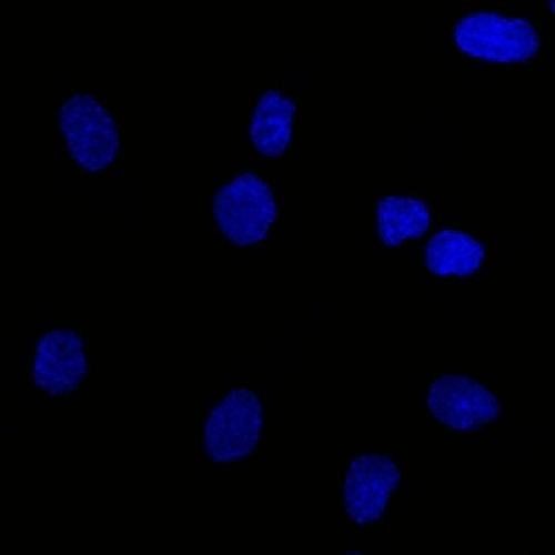
Confocal immunofluorescence of human colorectal carcinoma. EpCAM-Monospecific Mouse Monoclonal Antibody (EGP40/1372) labeled with CF488 (green); Nuclei are labeled with Reddot (red).

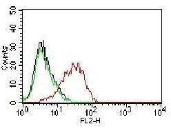
Flow Cytometric Analysis of PFA-fixed MCF-7 cells using EpCAM-Monospecific Mouse Monoclonal Antibody (EGP40/1372) followed by goat anti-mouse IgG-CF488 (blue); isotype control (red).

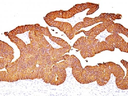
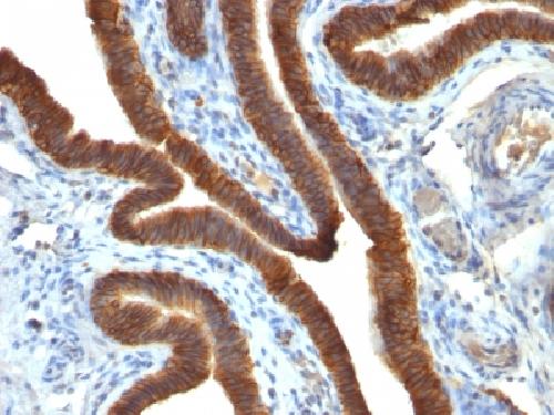
Formalin-fixed, paraffin-embedded human colorectal carcinoma stained with EpCAM-Monospecific Mouse Monoclonal Antibody (EGP40/1372).

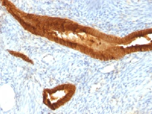
Formalin-fixed, paraffin-embedded human hepatocellular carcinoma stained with EpCAM-Monospecific Mouse Monoclonal Antibody (EGP40/1372).
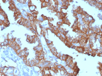
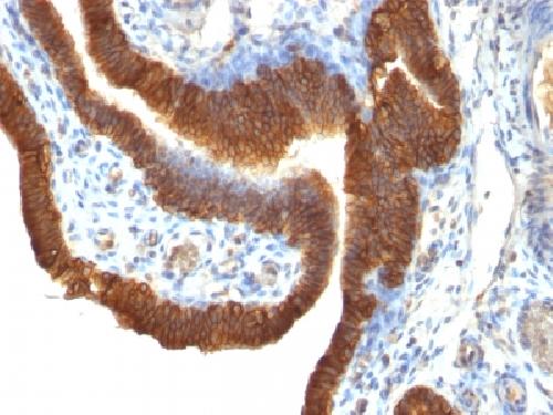
Formalin-fixed, paraffin-embedded Human Ovarian Carcinoma stained with EpCAM Mouse Monoclonal Antibody (EGP40/1372) conjugated to HRP.
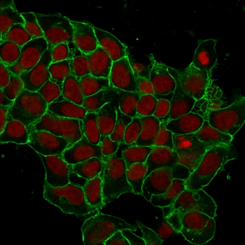
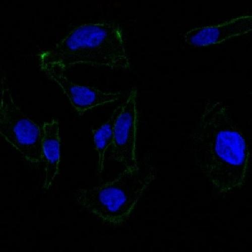
Immunofluorescence analysis of MCF-7 cells. EpCAM-Monospecific Mouse Monoclonal Antibody (EGP40/1372) labeled with CF488 (green). Nuclei are stained with RedDot (red).

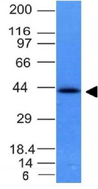
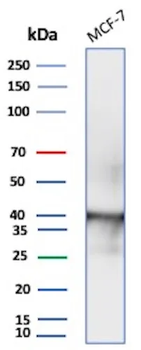
Western Blot Analysis of MCF-7 lysate using EpCAM Mouse Monoclonal Antibody (EGP40/1372).

Western Blot Analysis of MCF-7 lysate using EpCAM Mouse Monoclonal Antibody (EGP40/1372).
Documents Download
Datasheet
Product Information
Request a Document
Protocol Information
WB
Western Blot (IB, immunoblot)
IHC
Immunohistochemistry
FC
Flow Cytometry
ELISA
Enzyme-linked Immunosorbent Assay (EIA)
Ep-CAM / CD326 (Extracellular Domain) (Epithelial Marker) Antibody (orb1410486)

- 0.0
Based on 0 reviews
Participating in our Biorbyt product reviews program enables you to support fellow scientists by sharing your firsthand experience with our products.
Login to Submit a ReviewAvailable Sizes
Select a size below