You have no items in your shopping cart.
Rabbit anti-EpCAM Recombinant Monoclonal Antibody
SKU: orb1519809
Description
Images & Validation
−Item 1 of 3
| Tested Applications | FC, ICC, IHC, IP, WB |
|---|---|
| Dilution range | WB - 1:1000; IP - 12 µl/1 mg lysate; IHC - 1:100 to 1:500. Epitope retrieval with citrate buffer pH6.0 is recommended for FFPE tissue sections.; ICC - 1:100 to 1:500. Epitope retrieval with citrate buffer pH6.0 is recommended for FFPE cell sections. |
| Reactivity | Human |
| Application Notes |
Key Properties
−| Antibody Type | Primary Antibody |
|---|---|
| Host | Rabbit |
| Clonality | Recombinant |
| Clone No. | BLR077G |
| Immunogen | Between 264 and C-terminus |
| Target | EpCAM |
Storage & Handling
−| Storage | 2 - 8°C |
|---|---|
| Form/Appearance | Whole IgG |
| Buffer/Preservatives | Borate Buffered Saline (BBS) pH 8.2 with 0.1% rAlbumin and 0.09% Sodium Azide |
| Concentration | 500 µg/ml |
| Disclaimer | For research use only |
Alternative Names
−HNPCC8; human epithelial glycoprotein-2; KS 1/4 antigen; KS1/4; KSA; M4S1; membrane component, chromosome 4, surface marker (35kD glycoprotein); MK-1; TACSTD1; TROP1; hEGP314; major gastrointestinal tumor-associated protein GA733-2; tumor-associated calcium signal transducer 1; EGP; ESA; MIC18; adenocarcinoma-associated antigen; CD antigen CD326; DIAR5; EGP-2; EGP314; EGP40; Ep-CAM; epithelial cell adhesion molecule; Epithelial cell surface antigen; Epithelial glycoprotein; epithelial glycoprotein 314; cell surface glycoprotein Trop-1
Similar Products
−- Item 1 of 3
Rabbit anti-EpCAM Recombinant Monoclonal Antibody [orb1519811]
FC, ICC, IHC, IP, WB
Human, Mouse
Rabbit
Recombinant
Unconjugated
100 μl - Item 1 of 1
Recombinant EpCAM / CD326 (Epithelial Marker) Antibody [orb389164]
FC, IF, IHC, WB
Human, Mouse
Rabbit
Monoclonal
Unconjugated
100 μg (without BSA and Azide), 100 μg, 20 μg - Item 1 of 1
Recombinant Ep-CAM / CD326 (Extracellular Domain) (Epithelial Marker) Antibody [orb389165]
FC, IF, IHC
Human, Mouse
Rabbit
Monoclonal
Unconjugated
100 μg (without BSA and Azide), 100 μg, 20 μg - Item 1 of 1
Rabbit anti-EpCAM Recombinant Monoclonal Antibody [orb1519504]
FC, ICC, IHC, IP, WB
Human
Rabbit
Recombinant
Unconjugated
10 μl - Item 1 of 1
Rabbit anti-EpCAM Recombinant Monoclonal Antibody [orb1519506]
FC, ICC, IHC, IP, WB
Human
Rabbit
Recombinant
Unconjugated
100 μl


Quality Guarantee
Explore bioreagents carefree to elevate your research. All our products are rigorously tested for performance. If a product does not perform as described on its datasheet, our scientific support team will provide expert troubleshooting, a prompt replacement, or a refund. For full details, please see our Terms & Conditions and Buying Guide. Contact us at [email protected].

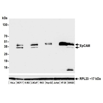
Detection of human EpCAM by western blot. Samples: Whole cell lysate (50 µg) from HeLa, MCF-7, K-562, LNCaP, RKO, Hep-G2, Jurkat, HT-29, and SW620 cells prepared using NETN lysis buffer. Antibody: Rabbit anti-EpCAM recombinant monoclonal antibody (orb1519809) used at 1:1000. Secondary: HRP-conjugated goat anti-rabbit IgG. Detection: Chemiluminescence with an exposure time of 10 seconds. Lower Panel: Rabbit anti-RPL23.
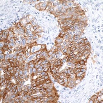
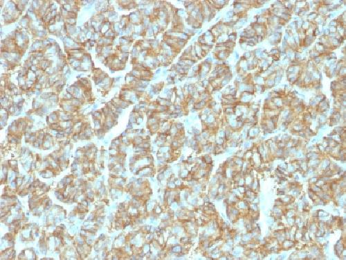
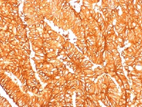
Detection of human EpCAM by immunohistochemistry. Sample: FFPE section of human ovarian carcinoma. Antibody: Rabbit anti-EpCAM recombinant monoclonal antibody (orb1519809) used at 1:100. Secondary: HRP-conjugated goat anti-rabbit IgG. Substrate: DAB.

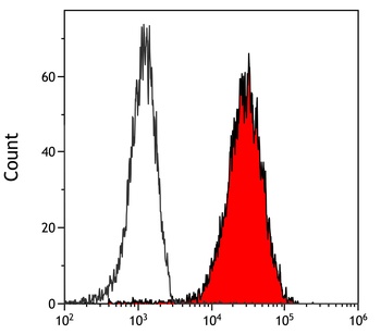
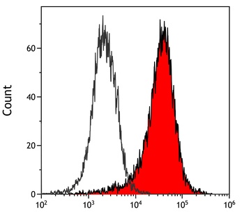
Detection of human EpCAM (shaded) in HT-29 cells by flow cytometry. Antibody: Rabbit anti-EpCAM recombinant monoclonal antibody (orb1519809) or isotype control (unshaded). Secondary: DyLight® 488-conjugated goat anti-rabbit IgG.
Quick Database Links
UniProt Details
− No UniProt data available
NCBI Reference Sequences
−Associated Accession Numbers
Curated reference sequences for the gene transcript and protein product| Protein | NP_006197.1 |
|---|
Documents Download
Datasheet
Product Information
Request a Document
Protocol Information
WB
Western Blot (IB, immunoblot)
IHC
Immunohistochemistry
FC
Flow Cytometry
ICC
Immunocytochemistry
IP
Immunoprecipitation
Rabbit anti-EpCAM Recombinant Monoclonal Antibody (orb1519809)

- 0.0
Based on 0 reviews
Participating in our Biorbyt product reviews program enables you to support fellow scientists by sharing your firsthand experience with our products.
Login to Submit a ReviewAvailable Sizes
Select a size below