You have no items in your shopping cart.
Featured
Description
Research Area
Epigenetics
Images & Validation
−Item 1 of 5
| Tested Applications | ChIP, EMSA, IF, IHC, WB |
|---|---|
| Dilution Range | WB: 1-500-1-1000, IHC-P: 1-100-1-200, IF/ICC: 1-100-1-500, ChIP: 1-100-1-500 |
| Reactivity | Gallus, Human, Mouse, Porcine, Rat, Sheep |
Key Properties
−| Antibody Type | Primary Antibody |
|---|---|
| Host | Rabbit |
| Clonality | Polyclonal |
| Immunogen | KLH-conjugated synthetic peptide encompassing a sequence within the C-term region of human AP2 alpha/beta. The exact sequence is proprietary. |
| Target | TFAP2A; TFAP2B |
| Purification | The antibody was purified by immunogen affinity chromatography. |
| Conjugation | Unconjugated |
Storage & Handling
−| Storage | Maintain refrigerated at 2-8°C for up to 2 weeks. For long term storage store at -20°C in small aliquots to prevent freeze-thaw cycles. |
|---|---|
| Form/Appearance | Liquid |
| Buffer/Preservatives | 0.42% Potassium phosphate, 0.87% Sodium chloride, pH 7.3, 30% glycerol, and 0.01% sodium azide. |
| Expiration Date | 12 months from date of receipt. |
| Disclaimer | For research use only |
Alternative Names
−TFAP2A; AP2TF; TFAP2; Transcription factor AP-2-alpha; AP2-alpha; AP-2 transcription factor; Activating enhancer-binding protein 2-alpha; Activator protein 2; AP-2; TFAP2B; Transcription factor AP-2-beta; AP2-beta; Activating enhancer-binding protein 2-beta
Similar Products
−
AP2 alpha + beta Rabbit Polyclonal Antibody (HRP) [orb478847]
ELISA, IHC-Fr, IHC-P
Canine, Equine, Gallus, Mouse, Porcine, Rabbit, Rat, Sheep
Human
Rabbit
Polyclonal
HRP
100 μlAP2 alpha + beta Rabbit Polyclonal Antibody [orb155674]
ELISA, ICC, IF, IHC-Fr, IHC-P
Canine, Equine, Gallus, Mouse, Porcine, Rabbit, Rat, Sheep
Human
Rabbit
Polyclonal
Unconjugated
50 μl, 100 μl, 200 μlAP2 alpha + beta Rabbit Polyclonal Antibody (Biotin) [orb457013]
ELISA, ICC, IF, IHC-Fr, IHC-P
Canine, Equine, Gallus, Mouse, Porcine, Rabbit, Rat, Sheep
Human
Rabbit
Polyclonal
Biotin
100 μlAP2 alpha + beta Rabbit Polyclonal Antibody (FITC) [orb465627]
ICC, IF
Canine, Equine, Gallus, Mouse, Porcine, Rabbit, Rat, Sheep
Human
Rabbit
Polyclonal
FITC
100 μlAP2 alpha + beta Rabbit Polyclonal Antibody (Cy5) [orb920621]
ICC, IF
Canine, Equine, Gallus, Mouse, Porcine, Rabbit, Rat, Sheep
Human
Rabbit
Polyclonal
Cy5
100 μl
Quality Guarantee
Explore bioreagents carefree to elevate your research. All our products are rigorously tested for performance. If a product does not perform as described on its datasheet, our scientific support team will provide expert troubleshooting, a prompt replacement, or a refund. For full details, please see our Terms & Conditions and Buying Guide. Contact us at [email protected].
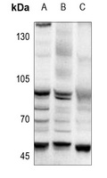
Western blot analysis of AP2 alpha/beta expression in PC3 (A), MCF7 (B), Myla2059 (C) whole cell lysates. (Predicted band size: 48; 50 kD; Observed band size: 48 kD)
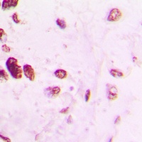
Immunohistochemical analysis of AP2 alpha/beta staining in human lung cancer formalin fixed paraffin embedded tissue section. The section was pre-treated using heat mediated antigen retrieval with sodium citrate buffer (pH 6.0). The section was then incubated with the antibody at room temperature and detected using an HRP conjugated compact polymer system. DAB was used as the chromogen. The section was then counterstained with haematoxylin and mounted with DPX.
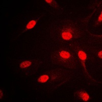
Immunofluorescent analysis of AP2 alpha/beta staining in HepG2 cells. Formalin-fixed cells were permeabilized with 0.1% Triton X-100 in TBS for 5-10 minutes and blocked with 3% BSA-PBS for 30 minutes at room temperature. Cells were probed with the primary antibody in 3% BSA-PBS and incubated overnight at 4 °C in a humidified chamber. Cells were washed with PBST and incubated with a DyLight 594-conjugated secondary antibody (red) in PBS at room temperature in the dark.
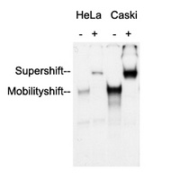
Anti-AP2 alpha/beta Antibody was used in an Electrophoretic Mobility Shift Assay (EMSA) to supershift the protein-DNA complex. Radiolabelled, double-stranded DNA oligonucleotides (10.000 cpm per lane) harbouring a binding site for AP2 alpha/beta were incubated with each 2 ug of nuclear extract (NE) from HeLa and Caski cells, respectively. Samples were incubated for 30 minutes at room temperature to allow the formation of protein-DNA complexes. Anti-AP2 alpha/beta Antibody were added to the samples (as indicated) and incubated for further 60 minutes at 4°C. Samples were separated in a 5.5% PAGE. The Gel was dried under vacuum and for autoradiography a X-ray film was exposed with an intensifying screen for 2 days at -80°C. Specific protein-DNA complexes were quantitatively supershifted with Anti-AP2 alpha/beta Antibody, verifying the binding of AP2 alpha/beta to the DNA oligonucleotide.
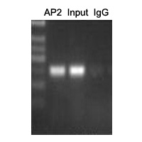
ChIP analysis of Cervical cancer cell lines lysate, incubated for 12 hours at 4°C. Cross-linking (X-ChIP) using formaldehyde for 10 minutes. Detection step: Semiquantitative PCR. Positive control: Tumor cell lines Hela. Negative control: Human primary keratinocytes.
Documents Download
Datasheet
Product Information
Request a Document
Protocol Information
WB
Western Blot (IB, immunoblot)
IHC
Immunohistochemistry
IF
Immunofluorescence
ChIP
Chromatin Immunoprecipitation
AP2 alpha/beta Rabbit Polyclonal Antibody (orb214653)

- 0.0
Based on 0 reviews
Participating in our Biorbyt product reviews program enables you to support fellow scientists by sharing your firsthand experience with our products.
Login to Submit a ReviewAvailable Sizes
Select a size below
Free Secondary Antibody (20 ul)0/0
Please add an antibody product to your cart first.