You have no items in your shopping cart.
Cpn10 Antibody
SKU: orb1822414
Description
Research Area
Cancer Biology, Cell Biology, Metabolism Research, Protein Biochemistry, Signal Transduction
Images & Validation
−Item 1 of 2
| Tested Applications | ELISA, ICC, IF, IHC, IP, WB |
|---|---|
| Dilution range | WB (1:1000), IHC (1:100); optimal dilutions for assays should be determined by the user. |
| Reactivity | Bovine, Canine, Frog, Guinea pig, Human, Mouse, Porcine, Rabbit, Rat, Sheep |
| Application Notes |
Key Properties
−| Host | Rabbit |
|---|---|
| Clonality | Polyclonal |
| Immunogen | Human Cpn10 peptide AA 91-101 |
| Target | Cpn10 |
| Molecular Weight | 10kDa |
| Purification | Protein A Purified |
| Conjugation | Unconjugated |
Storage & Handling
−| Storage | Maintain refrigerated at 2-8°C for up to 2 weeks. For long term storage store at -20°C in small aliquots to prevent freeze-thaw cycles. |
|---|---|
| Buffer/Preservatives | PBS pH 7.3, 0.02% sodium azide *Storage buffer changes when conjugated |
| Concentration | 1 mg/ml |
| Disclaimer | For research use only |
Alternative Names
−10kDa Chaperonin, Chaperonin 10, Cpn 10, EPF, GROES, Heat Shock 10kD protein1, HSP10, HSPE1
Similar Products
−- Item 1 of 4
HSP10 rabbit pAb Antibody [orb768669]
ELISA, IF, IHC, WB
Human, Mouse, Rat
Polyclonal
Unconjugated
100 μl, 50 μl - Item 1 of 1
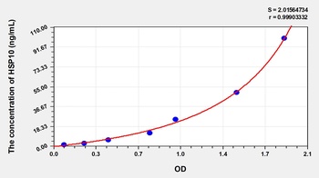
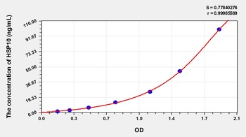
Mouse Heat Shock 10kDa Protein 1 (HSP10) ELISA Kit [orb777463]
Mouse
1.57-100 ng/mL
0.62 ng/mL
48 T, 96 T - Item 1 of 1
Human Heat Shock 10kDa Protein 1 (HSP10) ELISA Kit [orb775918]
Human
1.57-100 ng/mL
0.57 ng/mL
96 T, 48 T - Item 1 of 4
- Item 1 of 4
Cpn10/HSPE1 Antibody [orb76303]
FC, ICC, IF, IHC, WB
Human, Mouse, Rat
Rabbit
Polyclonal
Unconjugated
100 μg


Quality Guarantee
Explore bioreagents carefree to elevate your research. All our products are rigorously tested for performance. If a product does not perform as described on its datasheet, our scientific support team will provide expert troubleshooting, a prompt replacement, or a refund. For full details, please see our Terms & Conditions and Buying Guide. Contact us at [email protected].
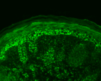
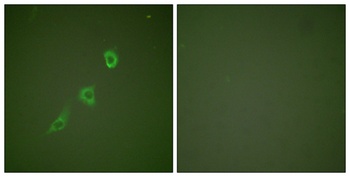
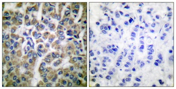
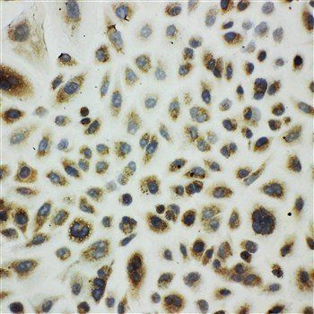
Immunohistochemistry analysis using Rabbit Anti-Cpn10 Polyclonal Antibody. Tissue: backskin. Species: Mouse. Fixation: Bouin's Fixative Solution. Primary Antibody: Rabbit Anti-Cpn10 Polyclonal Antibody at 1:100 for 1 hour at RT. Secondary Antibody: FITC Goat Anti-Rabbit (green) at 1:50 for 1 hour at RT. Localization: Strong basal layer punctate staining in the cytoplasm, weakens as it ascends to upper epidermis.
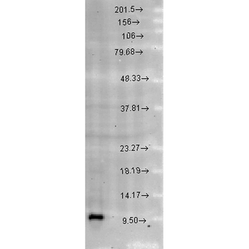
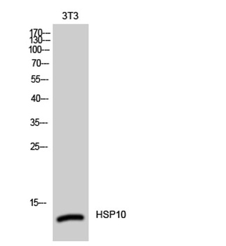
Western blot analysis of Rat brain cell lysates showing detection of Cpn10 protein using Rabbit Anti-Cpn10 Polyclonal Antibody. Load: 15 μgprotein. Block: 1.5% BSA for 30 minutes at RT. Primary Antibody: Rabbit Anti-Cpn10 Polyclonal Antibody at 1:1000 for 2 hours at RT. Secondary Antibody: Donkey Anti-Rabbit IgG: HRP for 1 hour at RT.
Quick Database Links
UniProt Details
− No UniProt data available
NCBI Gene Details
− No NCBI Gene data available
NCBI Reference Sequences
−Associated Accession Numbers
Curated reference sequences for the gene transcript and protein product| Protein | NP_002148.1 |
|---|
Documents Download
Datasheet
Product Information
Request a Document
Protocol Information
WB
Western Blot (IB, immunoblot)
IHC
Immunohistochemistry
IF
Immunofluorescence
ICC
Immunocytochemistry
ELISA
Enzyme-linked Immunosorbent Assay (EIA)
IP
Immunoprecipitation
Cpn10 Antibody (orb1822414)

- 0.0
Based on 0 reviews
Participating in our Biorbyt product reviews program enables you to support fellow scientists by sharing your firsthand experience with our products.
Login to Submit a ReviewAvailable Sizes
Select a size below