You have no items in your shopping cart.
Rabbit anti-Lamin-A/C Antibody
SKU: orb1524852
Description
Images & Validation
−Item 1 of 4
| Tested Applications | ICC, IHC, IP, WB |
|---|---|
| Dilution range | WB - 1:2,000 - 1:10,000; IP - 2 - 10 µg/mg lysate; IHC - 1:200 - 1:1,000. Epitope retrieval with citrate buffer pH 6.0 is recommended for FFPE tissue sections.; ICC-IF - 1:100 - 1:500. Formaldehyde fixation is recommended. Permeabilization with Triton-X 100 is recommended for formaldehyde-fixed cells. |
| Reactivity | Human, Mouse |
| Application Notes |
Key Properties
−| Antibody Type | Primary Antibody |
|---|---|
| Host | Rabbit |
| Clonality | Polyclonal |
| Immunogen | Between 1 and 50 |
| Target | Lamin-A/C |
| Conjugation | Unconjugated |
Storage & Handling
−| Storage | 2 - 8°C |
|---|---|
| Form/Appearance | Whole IgG |
| Buffer/Preservatives | Tris-buffered Saline containing 0.1% rAlbumin and 0.09% Sodium Azide |
| Concentration | 200 µg/ml |
| Disclaimer | For research use only |
Alternative Names
−prelamin-A/C; LDP1; LGMD1B; LMN1; LMNC; LMNL1; lamin; mandibuloacral dysplasia type A; lamin A/C-like 1; PRO1; renal carcinoma antigen NY-REN-32; MADA; CDCD1; LFP; 70 kDa lamin; IDC; CDDC; CMD1A; CMT2B1; epididymis secretory sperm binding protein; FPL; FPLD; FPLD2; HGPS; EMD2
Similar Products
−- Item 1 of 4
Rabbit anti-Lamin-A/C Antibody [orb1524853]
ICC, IHC, IP, WB
Human, Mouse
Rabbit
Polyclonal
Unconjugated
20 μg - Item 1 of 2
Lamin A/C LMNA Rabbit Monoclonal Antibody [orb547317]
FC, ICC, IF, IHC, IP, WB
Human
Rabbit
Monoclonal
Unconjugated
100 μl - Item 1 of 1
- Item 1 of 1
- Item 1 of 1
Lamin A/C Rabbit Monoclonal Antibody [orb866297]
FC, ICC, IF, IHC, WB
Human, Mouse, Rat
Rabbit
Monoclonal
Unconjugated
100 μl


Quality Guarantee
Explore bioreagents carefree to elevate your research. All our products are rigorously tested for performance. If a product does not perform as described on its datasheet, our scientific support team will provide expert troubleshooting, a prompt replacement, or a refund. For full details, please see our Terms & Conditions and Buying Guide. Contact us at [email protected].
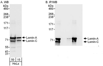
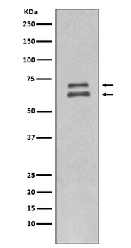
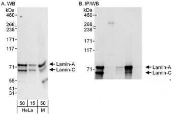
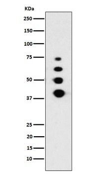
Detection of human Lamin-A and Lamin-C by western blot and immunoprecipitation. Samples: Whole cell lysate (15 and 50 µg for WB; 1 mg for IP, 20% of IP loaded) from HeLa cells. Antibodies: Affinity purified rabbit anti-Lamin-A/C antibody orb1524852 used for WB at 0.04 µg/ml (A) and 0.4 µg/ml (B) and used for IP at 6 µg/mg lysate. Lamin-A was also immunoprecipitated by rabbit anti-Lamin-A antibody, which recognizes a downstream epitope. Detection: Chemiluminescence with exposure times of 1 second (A and B).

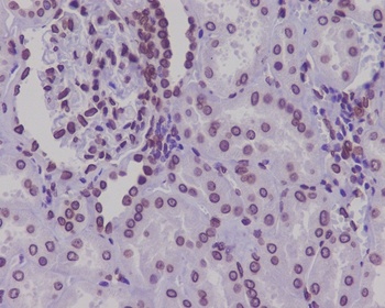
Detection of mouse Lamin-A /C by immunohistochemistry.Sample: FFPE section of mouse teratoma. Antibody: Affinity purified rabbit anti-Lamin-A/C (Cat. No. orb1524852) used at a dilution of 1: 200 (1µg/ml). Detection: DAB
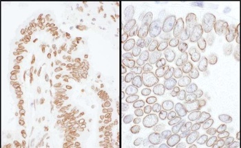
Detection of human Lamin-A /C by immunohistochemistry. Samples: FFPE sections of human colon carcinoma. Antibody: Affinity purified rabbit anti-Lamin-A/C (Cat. No. orb1524852) used at a dilution of 1: 200 (1µg/ml) (left) and 1: 1, 000 (0.2µg/ml) (right). Detection: DAB
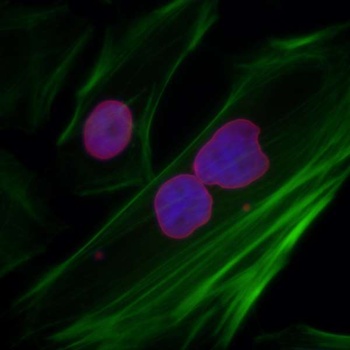
Detection of human Lamin-A /C by immunocytochemistry. Samples: Formaldehyde-fixed asynchronous HeLa cells. Antibody: Affinity purified rabbit anti-Lamin-A/C (Cat. No. orb1524852) used at a dilution of 1: 200 (1µg/ml). Detection: Red-fluorescent goat anti-rabbit IgG-heavy and light chain cross-adsorbed Antibody DyLight® 594 Conjugated used at a dilution of 1: 100.
Quick Database Links
UniProt Details
− No UniProt data available
NCBI Reference Sequences
−Associated Accession Numbers
Curated reference sequences for the gene transcript and protein product| Protein | NP_733821.1 |
|---|
Documents Download
Datasheet
Product Information
Request a Document
Protocol Information
WB
Western Blot (IB, immunoblot)
IHC
Immunohistochemistry
ICC
Immunocytochemistry
IP
Immunoprecipitation
Rabbit anti-Lamin-A/C Antibody (orb1524852)

- 0.0
Based on 0 reviews
Participating in our Biorbyt product reviews program enables you to support fellow scientists by sharing your firsthand experience with our products.
Login to Submit a ReviewAvailable Sizes
Select a size below