You have no items in your shopping cart.
p38 (pT180) Antibody
SKU: orb2651002
Description
Images & Validation
−Item 1 of 4
| Tested Applications | IF, IP, WB |
|---|---|
| Dilution Range | WB: WB (1/500 - 1/1000), IF/IC (1/100 - 1/500), IP (1/10 - 1/100), IF: WB (1/500 - 1/1000), IF/IC (1/100 - 1/500), IP (1/10 - 1/100), CHIP: WB (1/500 - 1/1000), IF/IC (1/100 - 1/500), IP (1/10 - 1/100), E: WB (1/500 - 1/1000), IF/IC (1/100 - 1/500), IP (1/10 - 1/100) |
| Reactivity | Human, Mouse, Rat, Zebrafish |
Key Properties
−| Host | Rabbit |
|---|---|
| Clonality | Polyclonal |
| Clone No. | MAPK14 |
| Conjugation | Unconjugated |
Storage & Handling
−| Storage | Maintain refrigerated at 2-8°C for up to 2 weeks. For long term storage store at -20°C in small aliquots to prevent freeze-thaw cycles. |
|---|---|
| Expiration Date | 12 months from date of receipt. |
| Disclaimer | For research use only |
Alternative Names
−CSBP; CSBP1; CSBP2; CSPB1; MXI2; SAPK2A; Mitogen-activated protein kinase 14; MAP kinase 14; MAPK 14; Cytokine suppressive anti-inflammatory drug-binding protein; CSAID-binding protein; CSBP; MAP kinase MXI2; MAX-interacting protein 2; Mitogen-activated protein kinase p38 alpha; MAP kinase p38 alpha; Stress-activated protein kinase 2a; SAPK2a
Similar Products
−- Item 1 of 2
p38 (pT180/Y182) Antibody [orb2650297]
IHC, IP, WB
Human, Mouse, Rat, Zebrafish
Rabbit
Polyclonal
Unconjugated
50 μl 


Quality Guarantee
Explore bioreagents carefree to elevate your research. All our products are rigorously tested for performance. If a product does not perform as described on its datasheet, our scientific support team will provide expert troubleshooting, a prompt replacement, or a refund. For full details, please see our Terms & Conditions and Buying Guide. Contact us at [email protected].
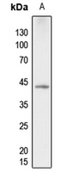
Western blot analysis of p38 (pT180) expression in zebrafish (A) whole cell lysates.
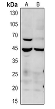
Western blot analysis of p38 (pT180) expression in mouse liver (A), rat liver (B) whole cell lysates.
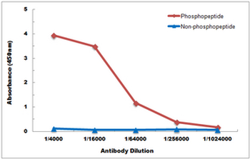
Direct ELISA antibody dose-response curve using Anti-p38 (pT180) Antibody. Antigen (phosphopeptide and non-phosphopeptide) concentration is 5 ug/ml. Goat Anti-Rabbit IgG (H&L) - HRP was used as the secondary antibody, and signal was developed by TMB substrate.
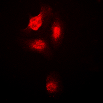
Immunofluorescent analysis of p38 (pT180) staining in HepG2 cells. Formalin-fixed cells were permeabilized with 0.1% Triton X-100 in TBS for 5-10 minutes and blocked with 3% BSA-PBS for 30 minutes at room temperature. Cells were probed with the primary antibody in 3% BSA-PBS and incubated overnight at 4°C in a humidified chamber. Cells were washed with PBST and incubated with a DyLight 594-conjugated secondary antibody (red) in PBS at room temperature in the dark.
Quick Database Links
UniProt
UniProt Details
− No UniProt data available
Documents Download
Datasheet
Product Information
Request a Document
Protocol Information
WB
Western Blot (IB, immunoblot)
IF
Immunofluorescence
IP
Immunoprecipitation
p38 (pT180) Antibody (orb2651002)

- 0.0
Based on 0 reviews
Participating in our Biorbyt product reviews program enables you to support fellow scientists by sharing your firsthand experience with our products.
Login to Submit a ReviewAvailable Sizes
Select a size below