You have no items in your shopping cart.
Description
Research Area
Neuroscience
Images & Validation
−Item 1 of 5
| Tested Applications | ELISA, FC, IF, IHC, WB |
|---|---|
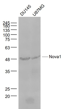
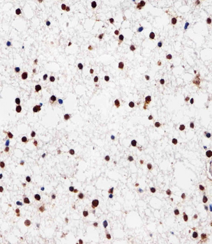
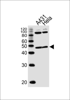
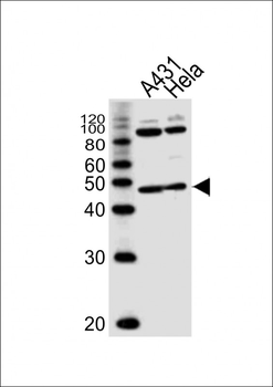
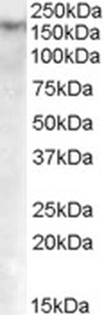
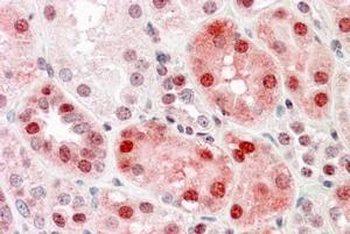
| Dilution Range | Peptide ELISA: antibody detection limit dilution 1:64000. Western blot: Approx. 50-55kDa band observed in Human Breast Cancer lysates, and in preliminary testing of Human Kidney lysate (calculated MW of 51.7kDa according to NP_002506.2). Recommended concentration: 0.01-0.03µg/ml. Primary incubation 1 hour at room temperature. IHC: Paraffin embedded Human Brain (Cortex) and Heart. Recommended concentration: 5µg/ml. Immunofluorescence: Strong expression of the protein seen in the nuclei of U2OS cells. Recommended concentration: 10µg/ml. Flow Cytometry: Flow cytometric analysis of U2OS cells. Recommended concentration: 10ug/ml. |
| Reactivity | Human |
Key Properties
−| Host | Goat |
|---|---|
| Clonality | Polyclonal |
| Target | NOVA1 |
| Protein Sequence | REMPQNVAKTEPVS |
| Molecular Weight | 51.7, 19.5 |
| Purification | Purified from goat serum by ammonium sulphate precipitation followed by antigen affinity chromatography using the immunizing peptide. |
| Conjugation | Unconjugated |
Storage & Handling
−| Storage | Maintain refrigerated at 2-8°C for up to 2 weeks. For long term storage store at -20°C in small aliquots to prevent freeze-thaw cycles. |
|---|---|
| Buffer/Preservatives | Supplied at 0.5 mg/ml in Tris saline, 0.02% sodium azide, pH 7.3 with 0.5% bovine serum albumin. Aliquot and store at -20°C. Minimize freezing and thawing. |
| Expiration Date | 12 months from date of receipt. |
| Disclaimer | For research use only |
Alternative Names
−anti NOVA1 antibody, anti neuro-oncological ventral antigen 1 antibody, anti Nova-1 antibody, anti paraneoplastic Ri antigen antibody, anti ventral neuron-specific protein 1 antibody
Similar Products
−- Item 1 of 12
Nova1 Rabbit Polyclonal Antibody [orb443203]
ELISA, FC, ICC, IF, IHC, IHC-Fr, WB
Human, Mouse, Rat
Rabbit
Polyclonal
Unconjugated
100 μg - Item 1 of 5
Nova1 Rabbit Polyclonal Antibody [orb158016]
IF, IHC-Fr, IHC-P, WB
Bovine, Gallus
Human, Mouse, Rat
Rabbit
Polyclonal
Unconjugated
100 μl, 200 μl, 50 μl - Item 1 of 3
NOVA1 Antibody (Center) [orb1926794]
IHC-P, WB
Mouse
Human, Mouse, Rat
Rabbit
Polyclonal
Unconjugated
100 μl, 50 μl - Item 1 of 2
NOVA1 Antibody (Center) [orb1926809]
IHC-P, WB
Mouse
Human, Mouse, Rat
Rabbit
Polyclonal
Unconjugated
100 μl, 50 μl - Item 1 of 4
Quality Guarantee
Explore bioreagents carefree to elevate your research. All our products are rigorously tested for performance. If a product does not perform as described on its datasheet, our scientific support team will provide expert troubleshooting, a prompt replacement, or a refund. For full details, please see our Terms & Conditions and Buying Guide. Contact us at [email protected].
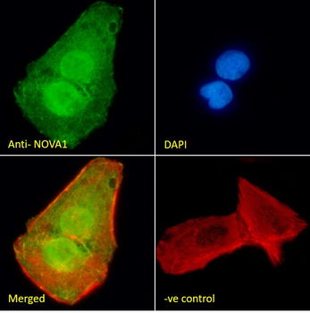
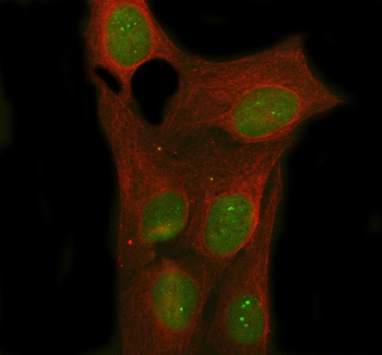
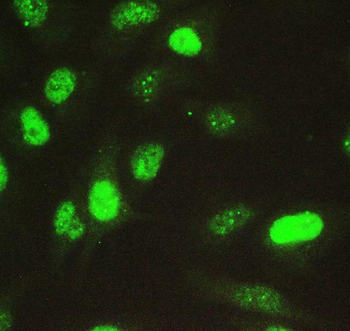
Immunofluorescence analysis of paraformaldehyde fixed U2OS cells, permeabilized with 0.15% Triton. Primary incubation 1hr (10 ug/ml) followed by Alexa Fluor 488 secondary antibody (2 ug/ml), showing nuclear staining. Actin filaments were stained with phalloidin (red) and the nuclear stain is DAPI (blue). Negative control: Unimmunized goat IgG (10 ug/ml) followed by Alexa Fluor 488 secondary antibody (2 ug/ml).
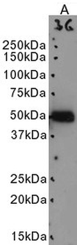
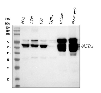
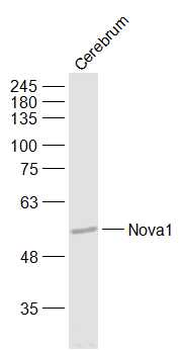
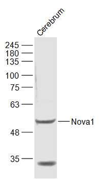
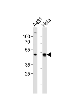
Primary incubation 1 hour at room temperature. Image A: Human Breast Cancer lysate at primary Ab concentration 0.03 µg/ml. (Loaded 35 µg protein in RIPA buffer, per lane). Detected by chemiluminescence.
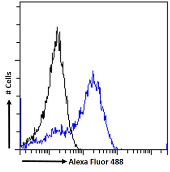
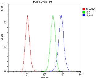
Flow cytometric analysis of paraformaldehyde fixed U2OS cells (blue line), permeabilized with 0.5% Triton. Primary incubation 1hr (10 ug/ml) followed by Alexa Fluor 488 secondary antibody (1 ug/ml). IgG control: Unimmunized goat IgG (black line) followed by Alexa Fluor 488 secondary antibody.
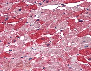
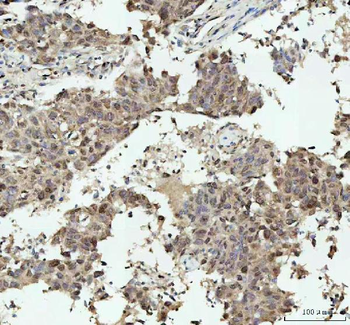
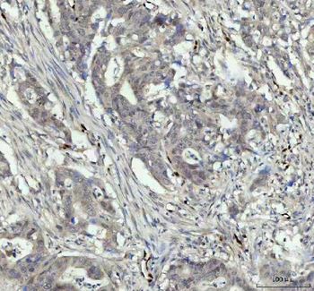
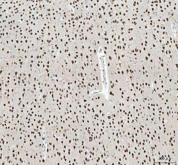
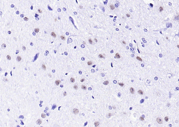
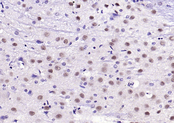
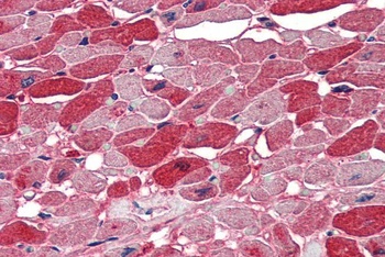
5 µg/ml staining of paraffin embedded Human Heart. Steamed antigen retrieval with citrate buffer pH6, AP-staining.
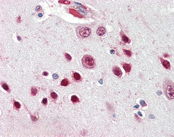
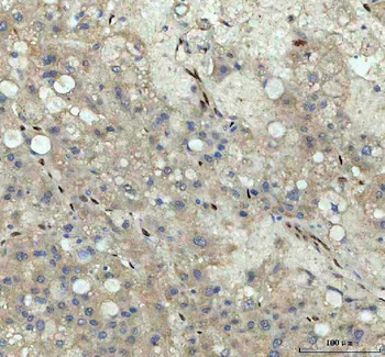
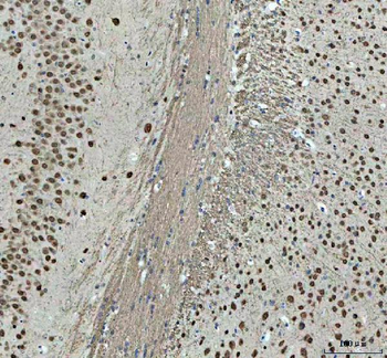
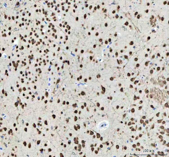
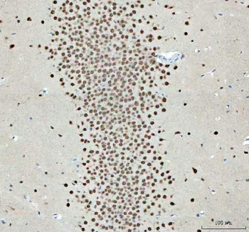
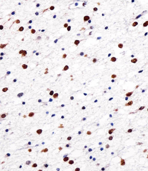
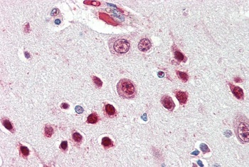
5 µg/ml staining of paraffin embedded Human Cortex. Steamed antigen retrieval with citrate buffer pH6, AP-staining.
Documents Download
Datasheet
Product Information
Request a Document
Protocol Information
WB
Western Blot (IB, immunoblot)
IHC
Immunohistochemistry
FC
Flow Cytometry
IF
Immunofluorescence
ELISA
Enzyme-linked Immunosorbent Assay (EIA)
NOVA1 Antibody (orb19796)

- 0.0
Based on 0 reviews
Participating in our Biorbyt product reviews program enables you to support fellow scientists by sharing your firsthand experience with our products.
Login to Submit a ReviewAvailable Sizes
Select a size below
Free Secondary Antibody (20 ul)0/0
Please add an antibody product to your cart first.