You have no items in your shopping cart.
Description
Research Area
Metabolism Research
Images & Validation
−Item 1 of 5
| Tested Applications | IF, IHC-P, WB |
|---|---|
| Dilution Range | WB - 1:1000, IF - 1:200, IHC-P - 1:100-500 |
| Reactivity | Human, Monkey, Mouse, Rat |
Key Properties
−| Antibody Type | Primary Antibody |
|---|---|
| Host | Rabbit |
| Clonality | Polyclonal |
| Isotype | Rabbit IgG |
| Molecular Weight | 57937 Da |
| Conjugation | Unconjugated |
Storage & Handling
−| Storage | Maintain refrigerated at 2-8°C for up to 2 weeks. For long term storage store at -20°C in small aliquots to prevent freeze-thaw cycles |
|---|---|
| Form/Appearance | Purified polyclonal antibody supplied in PBS with 0.09% (W/V) sodium azide. This antibody is prepared by Saturated Ammonium Sulfate (SAS) precipitation followed by dialysis against PBS. |
| Expiration Date | 12 months from date of receipt. |
| Disclaimer | For research use only |
Alternative Names
−OIP3, PK2, PK3, PKM2
Similar Products
−- Item 1 of 2
Quality Guarantee
Explore bioreagents carefree to elevate your research. All our products are rigorously tested for performance. If a product does not perform as described on its datasheet, our scientific support team will provide expert troubleshooting, a prompt replacement, or a refund. For full details, please see our Terms & Conditions and Buying Guide. Contact us at [email protected].
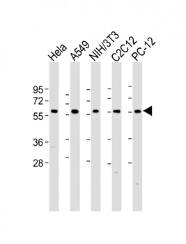
All lanes: Anti-hPKM2-N491 at 1:2000 dilution. Lane 1: Hela whole cell lysate. Lane 2: A549 whole cell lysate. Lane 3: NIH/3T3 whole cell lysate. Lane 4: C2C12 whole cell lysate. Lane 5: PC-12 whole cell lysate.Lysates/proteins at 20 µg per lane. Secondary Goat Anti-Rabbit IgG, (H+L), Peroxidase conjugated at 1/10000 dilution. Predicted band size: 58 kDa. Blocking/Dilution buffer: 5% NFDM/TBST.
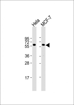
All lanes: Anti-PKM2 Antibody (N491) at 1:1000 dilution. Lane 1: Hela whole cell lysate. Lane 2: MCF-7 whole cell lysate.Lysates/proteins at 20 µg per lane. Secondary Goat Anti-Rabbit IgG, (H+L), Peroxidase conjugated at 1/10000 dilution. Predicted band size: 58 kDa. Blocking/Dilution buffer: 5% NFDM/TBST.
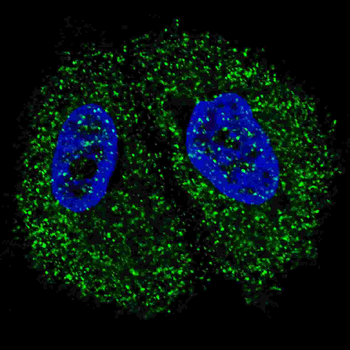
Fluorescent confocal image of MCF7 cells stained with Pyruvate Kinase (PKM2) (C-term) Antibody. MCF7 cells were fixed with 4% PFA (20 min), permeabilized with Triton X-100 (0.2%, 30 min). Cells were then incubated with Pyruvate Kinase (PKM2) (C-term) primary antibody (1:200, 2 h at room temperature). For secondary antibody, Alexa Fluor 488 conjugated donkey anti-rabbit antibody (green) was used (1:1000, 1h). Nuclei were counterstained with Hoechst 33342 (blue) (10 μg/ml, 5 min).
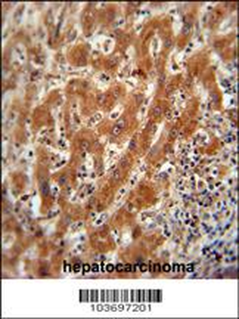
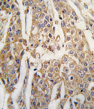
Pyruvate Kinase (PKM2) Antibody (C-term) immunohistochemistry analysis in formalin fixed and paraffin embedded human hepatocarcinoma followed by peroxidase conjugation of the secondary antibody and DAB staining. This data demonstrates the use of the Pyruvate Kinase (PKM2) Antibody (C-term) for immunohistochemistry. Clinical relevance has not been evaluated.
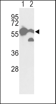
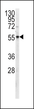
Western blot analysis of hPKM2-N491 in A2058 cell line (lane 1) and mouse brain tissue (lane 2) lysates (35 ug/lane). PKM2 (arrow) was detected using the purified Pab.
Quick Database Links
UniProt
UniProt Details
− No UniProt data available
Documents Download
Datasheet
Product Information
Request a Document
Protocol Information
WB
Western Blot (IB, immunoblot)
IHC-P
Immunohistochemistry Paraffin
IF
Immunofluorescence
Pyruvate Kinase (PKM2) Antibody (C-term) (orb1939499)

- 0.0
Based on 0 reviews
Participating in our Biorbyt product reviews program enables you to support fellow scientists by sharing your firsthand experience with our products.
Login to Submit a ReviewAvailable Sizes
Select a size below
Free Secondary Antibody (20 ul)0/0
Please add an antibody product to your cart first.