You have no items in your shopping cart.
Description
Research Area
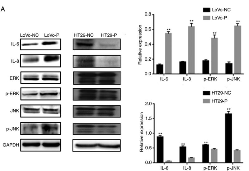
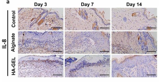
Immunology & Inflammation
Images & Validation
−
Item 1 of 2
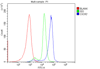
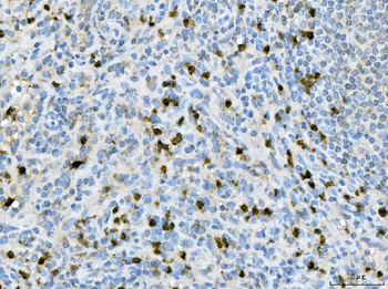
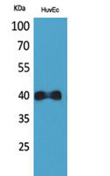
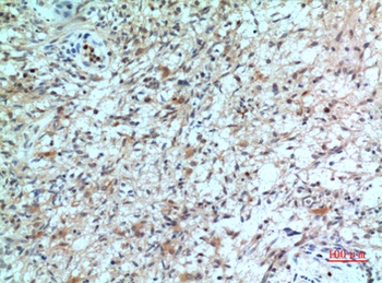
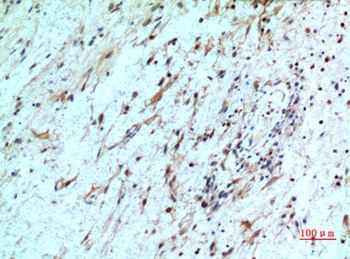
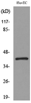
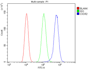
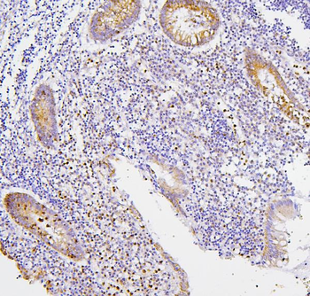
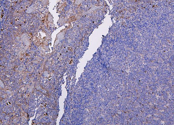
| Tested Applications | FC, IHC |
|---|---|
| Dilution Range | Immunohistochemistry (Paraffin-embedded Section), 0.5-1μg/ml, Human Flow Cytometry (Fixed), 1-3μg/1x10^6 cells, Human |
| Reactivity | Human |
Related Conjugates & Formulations
−Key Properties
−| Antibody Type | Primary Antibody |
|---|---|
| Host | Rabbit |
| Clonality | Polyclonal |
| Isotype | Rabbit IgG |
| Immunogen | A synthetic peptide corresponding to a sequence at the N-terminus of human CXCR2, which shares 52.4% amino acid (aa) sequence identity with rat CXCR2. |
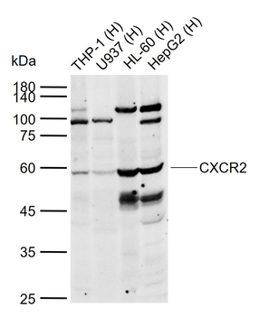
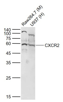
| Target | C-X-C chemokine receptor type 2 |
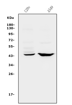
| Molecular Weight | 68 kDa |
| Purification | Immunogen affinity purified. |
| Conjugation | Unconjugated |
Storage & Handling
−| Storage | Maintain refrigerated at 2-8°C for up to 2 weeks. For long term storage store at -20°C in small aliquots to prevent freeze-thaw cycles. |
|---|---|
| Form/Appearance | Lyophilized |
| Buffer/Preservatives | Each vial contains 4 mg Trehalose, 0.9 mg NaCl and 0.2 mg Na2HPO4. |
| Concentration | 500 µg/ml |
| Expiration Date | 12 months from date of receipt. |
| Disclaimer | For research use only |
Alternative Names
−Neutrophil gelatinase-associated lipocalin; NGAL; Lipocalin-2; SV-40-induced 24P3 protein; Siderocalin LCN2; p25; Lcn2
Similar Products
−- Item 1 of 12
IL8 Antibody [orb229133]
IHC-P, WB
Human
Rabbit
Polyclonal
Unconjugated
- Item 1 of 2
CXCR2 Rabbit Polyclonal Antibody [orb1720]
WB
Rat
Human, Mouse
Rabbit
Polyclonal
Unconjugated
50 μl, 100 μl, 200 μl - Item 1 of 4
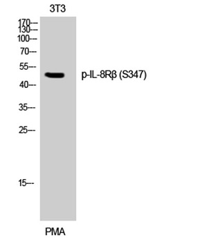
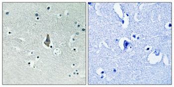
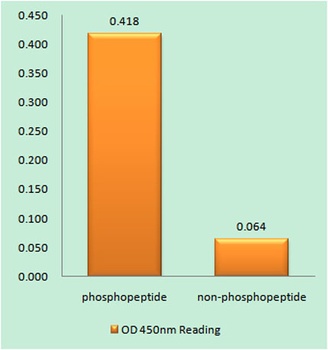
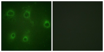
IL-8Rβ (phospho Ser347) rabbit pAb Antibody [orb768761]
ELISA, IF, IHC, WB
Human, Mouse
Polyclonal
Unconjugated
50 μl, 100 μl - Item 1 of 4
IL-8Rβ rabbit pAb Antibody [orb767002]
ELISA, IF, IHC, WB
Human, Mouse, Rat
Polyclonal
Unconjugated
50 μl, 100 μl - Item 1 of 4
CXCR2 Rabbit Polyclonal Antibody [orb669171]
ELISA, FC, IHC, WB
Human
Rabbit
Polyclonal
Unconjugated
100 μg
Quality Guarantee
Explore bioreagents carefree to elevate your research. All our products are rigorously tested for performance. If a product does not perform as described on its datasheet, our scientific support team will provide expert troubleshooting, a prompt replacement, or a refund. For full details, please see our Terms & Conditions and Buying Guide. Contact us at [email protected].
Quick Database Links
Gene Symbol
C-X-C chemokine receptor type 2
UniProt
UniProt Details
− No UniProt data available
Protocol Information
IHC
Immunohistochemistry
FC
Flow Cytometry
Available Sizes
Select a size below
Free Secondary Antibody (20 ul)0/0
Please add an antibody product to your cart first.