
You have no items in your shopping cart.
Secondary antibodies
Amplify your signals and achieve crisp, background-free detection. Explore Biorbyt's range of secondary antibodies, including highly cross-adsorbed options, fragment antibodies, and highly photostable fluorescent or enzymatic conjugates for western blot, IHC, ELISA, and flow cytometry.
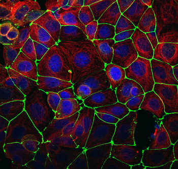
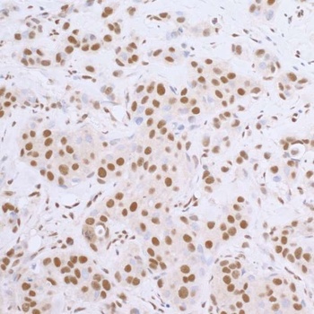
Cross-adsorbed
Chemically purified to minimize species cross-reactivity, these antibodies are essential for multiplex experiments where multiple primary host antibodies are used.
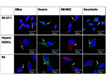
Fluorescent labels
Featuring bright, highly photostable fluorescent labels (FITC, DyLight, and Alexa Fluor equivalents) optimized for immunofluorescence and flow cytometry workflows.
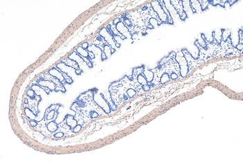
Enzymatic labels
High-purity HRP (Horseradish Peroxidase) and AP (Alkaline Phosphatase) conjugates optimized for high-sensitivity western blotting and colorimetric ELISA.
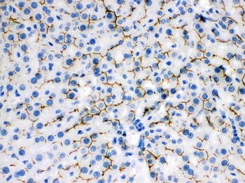
Biotinylated secondaries
Designed for high-affinity pairing with streptavidin complexes, biotin-conjugated secondary antibodies provide massive signal amplification for low-abundance targets.
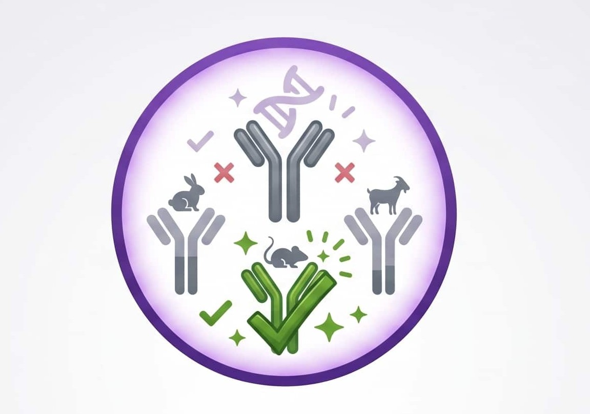
How to Choose the Right Secondary Antibody
Selecting the correct secondary antibody is vital to the success of your experiment. Your choice depends directly on the host species of your primary antibody, the primary antibody subclass, and your detection system.
Read our technical guide to learn how to navigate host species mismatching, cross-adsorption selection, and conjugation types.
Commitment to antibody specificity and reproducibility
At Biorbyt, we help researchers select secondaries with absolute confidence by combining quality-driven production with dedicated specificity strategies.
Our portfolio includes highly cross-adsorbed antibodies to prevent cross-reactivity in multi-labeling experiments, fragment antibodies to resolve Fc receptor background issues, and robust HRP/fluorescent labels for clear data publication.
We recognize that performance is application-dependent, so we focus on clear technical specifications to help you find the correct matching reagent for your assay design.
- High-specificity cross-adsorbed options for multiplexing assays
- F(ab')2 and Fab fragments to minimize non-specific Fc-receptor binding
- Highly photostable fluorescent conjugates (FITC, Alexa Fluor equivalents)
- HRP and AP enzymatic conjugates for western blot and IHC
Secondary antibodies are essential tools for amplifying signals by binding to primary antibodies targeted at specific proteins. Biorbyt offers a diverse selection covering common host species as well as specialized formats for advanced analytical setups.
The Essentials: Most Used Secondary Antibodies
Application Need | Product Description | Conjugate | SKU |
|---|---|---|---|
Routine WB & ELISA | Goat Anti-Rabbit IgG (H+L) Antibody (HRP) High sensitivity, broad species utility. | HRP | |
Routine WB & ELISA | Goat Anti-mouse IgG (H+L) Antibody (HRP) Generated against a Mouse IgG (H+L) | HRP | |
Routine WB & ELISA | Goat Anti-human IgG (H+L) Antibody (HRP) Generated against a Human IgG (H+L) | HRP | |
Multiplexing (Low Background) | Goat Anti-Rabbit IgG (H&L)- Polymer (Ready to use) Antibody (HRP) Cross-adsorbed against IgG from bovine, goat, guinea pig, hamster, horse, mouse, rat, and human. | HRP | |
Immunofluorescence / FC | Goat Anti-Rabbit IgG (H+L) FITC Bright, stable green fluorescence. | FITC | |
Signal Amplification | HRP Goat Rabbit IgG Fc Antibody Recognizes rabbit IgG F(c) fragment. | HRP |
F(ab')2 & Fab fragments
Enzymatically cleaved to remove the Fc region, these fragments prevent non-specific binding to tissue Fc receptors, drastically reducing background noise in lymphoid tissues.
Signal amplification
Use the remarkable binding affinity of biotin and streptavidin. This system offers massive signal multiplication for low-expression targets in IHC and ELISA.
Isotype controls
Isotype controls help distinguish non-specific background signal from actual antibody staining in flow cytometry and immunohistochemistry experiments.
VHH / Nanobodies
Single-domain antibodies derived from camelids. Their tiny size (~15 kDa) allows for exceptional tissue penetration and high-resolution imaging.