You have no items in your shopping cart.
Description
Research Area
Protein Biochemistry, Signal Transduction
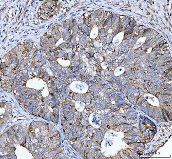
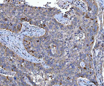
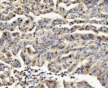
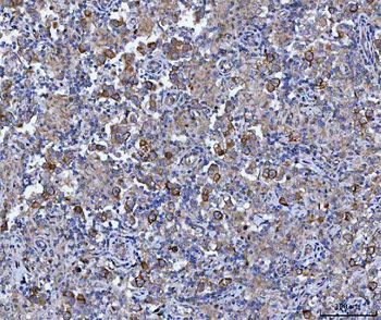
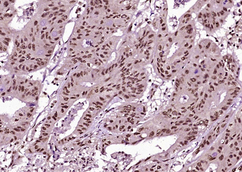
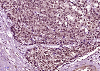
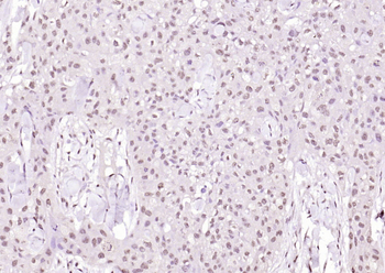
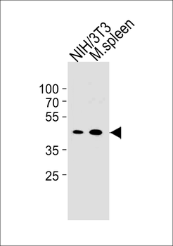
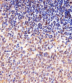
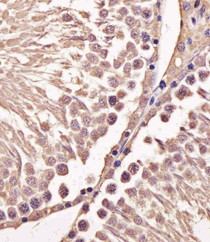
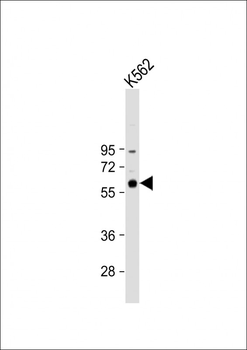
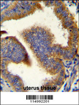
Images & Validation
−

Item 1 of 3
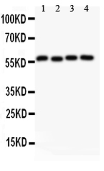
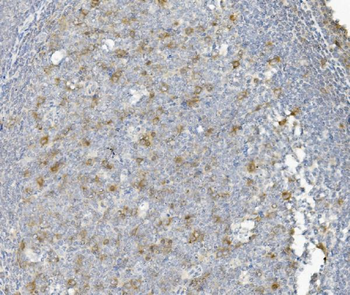
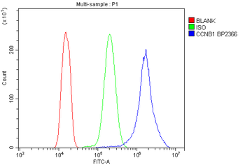
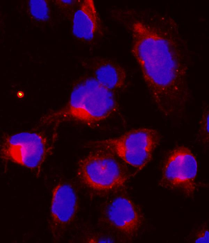
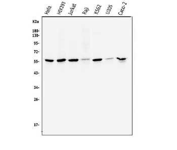
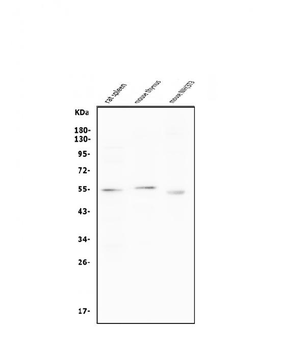
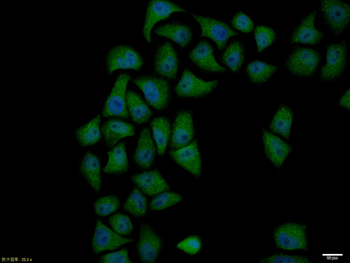
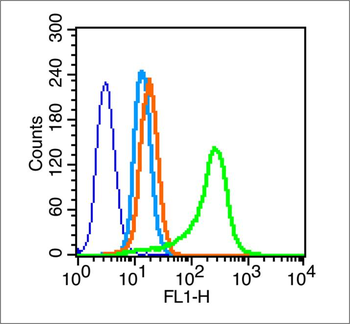
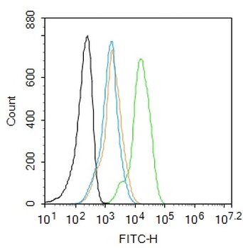
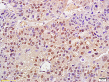
| Tested Applications | FC, ICC, IF, WB |
|---|---|
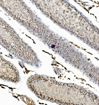
| Dilution Range | Western blot, 0.1-0.5μg/ml, Human Immunocytochemistry/Immunofluorescence, 2μg/ml, Human Flow Cytometry (Fixed), 1-3μg/1x10^6 cells, Human |
| Reactivity | Human |
Related Conjugates & Formulations
−Key Properties
−| Antibody Type | Primary Antibody |
|---|---|
| Host | Rabbit |
| Clonality | Polyclonal |
| Isotype | Rabbit IgG |
| Immunogen | E.coli-derived human Cyclin B1 recombinant protein (Position: M1-V433). Human Cyclin B1 shares 86% and 85% amino acid (aa) sequences identity with mouse and rat Cyclin B1, respectively. |
| Target | G2/mitotic-specific cyclin-B1 |
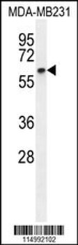
| Molecular Weight | 56 kDa |
| Purification | Immunogen affinity purified. |
| Conjugation | Unconjugated |
Storage & Handling
−| Storage | Maintain refrigerated at 2-8°C for up to 2 weeks. For long term storage store at -20°C in small aliquots to prevent freeze-thaw cycles. |
|---|---|
| Form/Appearance | Lyophilized |
| Buffer/Preservatives | Each vial contains antibody formulated with stabilizing components, 0.9 mg NaCl, 0.2 mg Na2HPO4, and 0.05 mg NaN3. *This antibody is supplied in a stabilized formulation. Compatibility with conjugation reactions depends on the chemistry of the conjugation method used. For conjugation methods that are not compatible with the stabilizing components present in this formulation, a carrier-free antibody format is required. |
| Concentration | Adding 0.2 ml of distilled water will yield a concentration of 500 μg/ml. |
| Expiration Date | 12 months from date of receipt. |
| Disclaimer | For research use only |
Alternative Names
−G2/mitotic-specific cyclin-B1; CCNB1; CCNB
Similar Products
−- Item 1 of 10
Cyclin B1/CCNB1 Rabbit Polyclonal Antibody [orb654315]
ELISA, FC, ICC, IF, IHC, WB
Human, Mouse, Rat
Rabbit
Polyclonal
Unconjugated
100 μg - Item 1 of 4
Phospho-Cyclin B1 (Ser126) Rabbit Polyclonal Antibody [orb5928]
ICC, IF, IHC-Fr, IHC-P
Human
Rabbit
Polyclonal
Unconjugated
50 μl, 100 μl, 200 μl - Item 1 of 3
M CCNB1 Antibody (Center S123/S125) [orb1927457]
IHC-P, WB
Hamster
Mouse
Rabbit
Polyclonal
Unconjugated
50 μl, 100 μl - Item 1 of 3
- Item 1 of 3
Phospho-Cyclin B1 (Ser147) Rabbit Polyclonal Antibody [orb5926]
FC, IF, IHC-Fr, IHC-P
Bovine, Canine, Equine, Gallus, Guinea pig, Mouse, Porcine, Rabbit, Rat
Human
Rabbit
Polyclonal
Unconjugated
50 μl, 100 μl, 200 μl
Quality Guarantee
Explore bioreagents carefree to elevate your research. All our products are rigorously tested for performance. If a product does not perform as described on its datasheet, our scientific support team will provide expert troubleshooting, a prompt replacement, or a refund. For full details, please see our Terms & Conditions and Buying Guide. Contact us at [email protected].
Quick Database Links
Gene Symbol
G2/mitotic-specific cyclin-B1
UniProt
UniProt Details
− No UniProt data available
Protocol Information
WB
Western Blot (IB, immunoblot)
FC
Flow Cytometry
IF
Immunofluorescence
ICC
Immunocytochemistry
Available Sizes
Select a size below
Free Secondary Antibody (20 ul)0/0
Please add an antibody product to your cart first.