You have no items in your shopping cart.
GPR54/KISS1R Rabbit Polyclonal Antibody (PE)
Description
Research Area
Images & Validation
−| Tested Applications | FC |
|---|---|
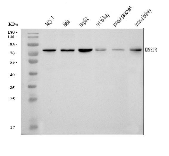
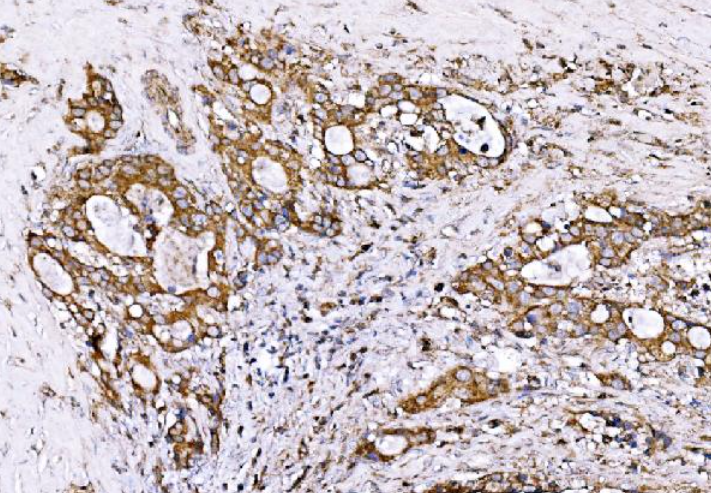
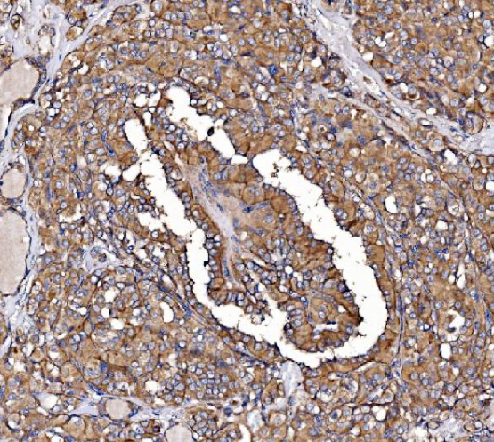
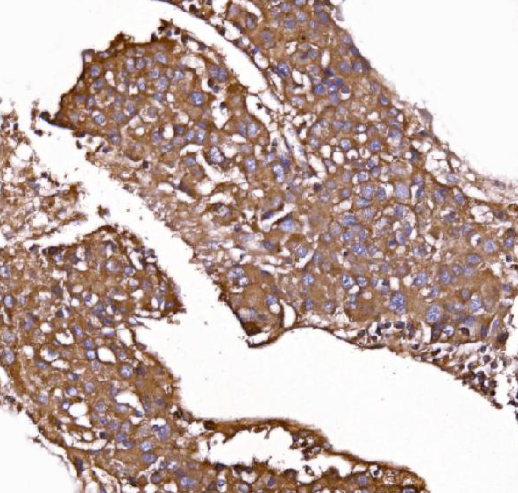
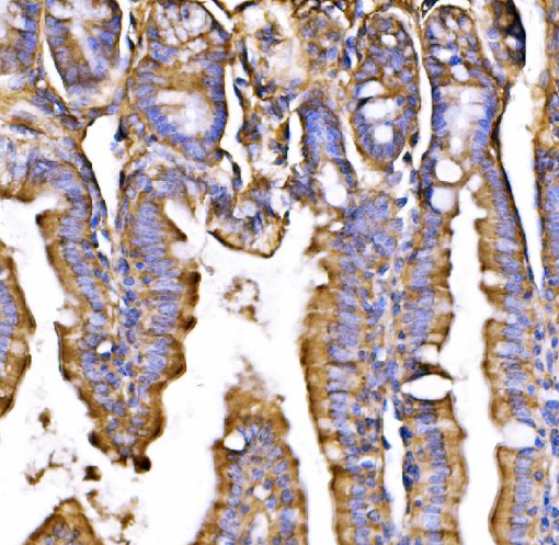
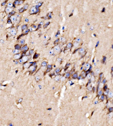
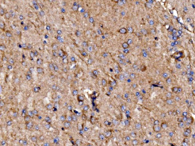
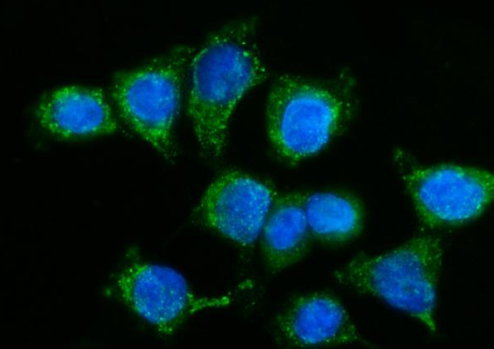
| Dilution Range | Western blot, 0.25-0.5 μg/ml, Immunohistochemistry(Paraffin-embedded Section), 1-2 μg/ml, Immunocytochemistry/Immunofluorescence, 5 μg/ml, ELISA, 0.1-0.5 μg/ml |
| Reactivity | Human, Mouse, Rat |
| Application Notes |
Key Properties
−| Antibody Type | Primary Antibody |
|---|---|
| Host | Rabbit |
| Clonality | Polyclonal |
| Isotype | Rabbit IgG |
| Immunogen | E.coli-derived human GPR54/KISS1R recombinant protein (Position: N60-A373). |
| Target | KiSS-1 receptor |
| Molecular Weight | Observed: 80 kDa |
| Purification | Immunogen affinity purified. |
| Conjugation | PE |
Storage & Handling
−| Storage | At -20°C for one year from date of receipt. Avoid repeated freezing and thawing. Protect from light. |
|---|---|
| Form/Appearance | Liquid |
| Buffer/Preservatives | Each vial contains 4 mg Trehalose, 0.9 mg NaCl, 0.2 mg Na2HPO4 |
| Expiration Date | 12 months from date of receipt. |
| Disclaimer | For research use only |
Alternative Names
−
Quality Guarantee
Explore bioreagents carefree to elevate your research. All our products are rigorously tested for performance. If a product does not perform as described on its datasheet, our scientific support team will provide expert troubleshooting, a prompt replacement, or a refund. For full details, please see our Terms & Conditions and Buying Guide. Contact us at [email protected].

Western blot analysis of GPR54/KISS1R using antiGPR54/KISS1R antibody. Electrophoresis was performed on a 5-20% SDS-PAGE gel at 70V (Stacking gel) / 90V (Resolving gel) for 2-3 hours. The sample well of each lane was loaded with 30 ug of sample under reducing conditions. Lane 1: human MCF-7 whole cell lysates, Lane 2: human Hela whole cell lysates, Lane 3: human HepG2 whole cell lysates, Lane 4: rat kindey tissue lysates, Lane 5: mouse pancreas tissue lysates, Lane 6: mouse kindey tissue lysates.

IHC analysis of GPR54/KISS1R using anti-GPR54/KISS1R antibody. GPR54/KISS1R was detected in a paraffin-embedded section of human gall bladder adenosquamous carcinoma tissue. Heat mediated antigen retrieval was performed in EDTA buffer (pH 8.0, epitope retrieval solution). The tissue section was blocked with 10% goat serum. The tissue section was then incubated with 2 μg/ml rabbit anti-GPR54/KISS1R Antibody overnight at 4°C. Biotinylated goat anti-rabbit IgG was used as secondary antibody and incubated for 30 minutes at 37°C. The tissue section was developed using Strepavidin-Biotin-Complex (SABC) with DAB as the chromogen

IHC analysis of GPR54/KISS1R using anti-GPR54/KISS1R antibody. GPR54/KISS1R was detected in a paraffin-embedded section of human hyroid papillary carcinoma tissue. Heat mediated antigen retrieval was performed in EDTA buffer (pH 8.0, epitope retrieval solution). The tissue section was blocked with 10% goat serum. The tissue section was then incubated with 2 μg/ml rabbit anti-GPR54/KISS1R Antibody overnight at 4°C. Biotinylated goat anti-rabbit IgG was used as secondary antibody and incubated for 30 minutes at 37°C. The tissue section was developed using Strepavidin-Biotin-Complex (SABC) with DAB as the chromogen

IHC analysis of GPR54/KISS1R using anti-GPR54/KISS1R antibody. GPR54/KISS1R was detected in a paraffin-embedded section of human liver cancer tissue. Heat mediated antigen retrieval was performed in EDTA buffer (pH 8.0, epitope retrieval solution). The tissue section was blocked with 10% goat serum. The tissue section was then incubated with 2 μg/ml rabbit anti-GPR54/KISS1R Antibody overnight at 4°C. Biotinylated goat anti-rabbit IgG was used as secondary antibody and incubated for 30 minutes at 37°C. The tissue section was developed using Strepavidin-Biotin-Complex (SABC) with DAB as the chromogen

IHC analysis of GPR54/KISS1R using anti-GPR54/KISS1R antibody. GPR54/KISS1R was detected in a paraffin-embedded section of mouse colon tissue. Heat mediated antigen retrieval was performed in EDTA buffer (pH 8.0, epitope retrieval solution). The tissue section was blocked with 10% goat serum. The tissue section was then incubated with 2 μg/ml rabbit anti-GPR54/KISS1R Antibody overnight at 4°C. Biotinylated goat anti-rabbit IgG was used as secondary antibody and incubated for 30 minutes at 37°C. The tissue section was developed using Strepavidin-Biotin-Complex with DAB as the chromogen

IHC analysis of GPR54/KISS1R using anti-GPR54/KISS1R antibody. GPR54/KISS1R was detected in a paraffin-embedded section of mouse brain tissue. Heat mediated antigen retrieval was performed in EDTA buffer (pH 8.0, epitope retrieval solution). The tissue section was blocked with 10% goat serum. The tissue section was then incubated with 2 μg/ml rabbit anti-GPR54/KISS1R Antibody overnight at 4°C. Biotinylated goat anti-rabbit IgG was used as secondary antibody and incubated for 30 minutes at 37°C. The tissue section was developed using Strepavidin-Biotin-Complex (SABC) with DAB as the chromogen

IHC analysis of GPR54/KISS1R using anti-GPR54/KISS1R antibody. GPR54/KISS1R was detected in a paraffin-embedded section of rat brain tissue. Heat mediated antigen retrieval was performed in EDTA buffer (pH 8.0, epitope retrieval solution). The tissue section was blocked with 10% goat serum. The tissue section was then incubated with 2 μg/ml rabbit anti-GPR54/KISS1R Antibody overnight at 4°C. Biotinylated goat anti-rabbit IgG was used as secondary antibody and incubated for 30 minutes at 37°C. The tissue section was developed using Strepavidin-Biotin-Complex (SABC) with DAB as the chromogen

IF analysis of GPR54/KISS1R using anti-GPR54/KISS1R antibody. GPR54/KISS1R was detected in an immunocytochemical section of CACO-2 cells. Enzyme antigen retrieval was performed using IHC enzyme antigen retrieval reagent for 15 mins. The cells were blocked with 10% goat serum. And then incubated with 5 μg/mL rabbit anti-GPR54/KISS1R Antibody overnight at 4°C. DyLight®488 Conjugated Goat Anti-Rabbit IgG was used as secondary antibody at 1:100 dilution and incubated for 30 minutes at 37°C. The section was counterstained with DAPI. Visualize using a fluorescence microscope and filter sets appropriate for the label used
Quick Database Links
UniProt Details
−Documents Download
Request a Document
GPR54/KISS1R Rabbit Polyclonal Antibody (PE) (orb2599870)
Participating in our Biorbyt product reviews program enables you to support fellow scientists by sharing your firsthand experience with our products.
Login to Submit a Review