You have no items in your shopping cart.
Description
Research Area
Cardiovascular Research, Cell Biology, Protein Biochemistry, Signal Transduction, Stem Cell & Developmental Biology
Images & Validation
−

Item 1 of 5
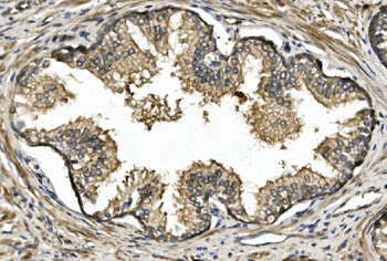
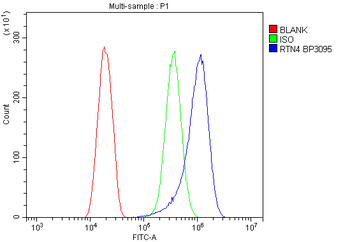
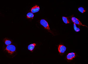
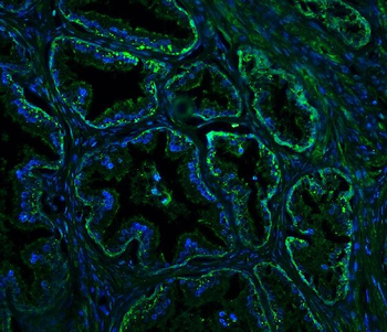
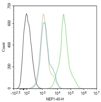
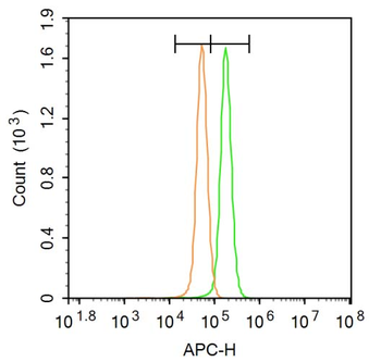
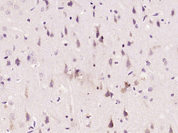
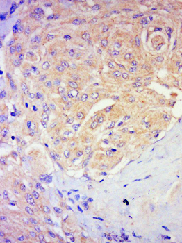
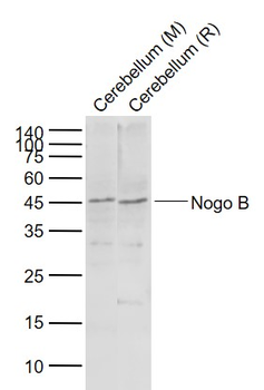
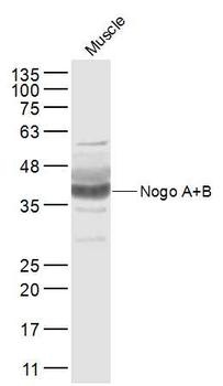
| Tested Applications | FC, ICC, IF, IHC, WB |
|---|---|
| Dilution Range | Western blot, 0.1-0.25μg/ml, Human Immunohistochemistry (Paraffin-embedded Section), 0.5-1μg/ml, human Immunocytochemistry/Immunofluorescence, 4μg/ml, Human Immunofluorescence, 4μg/ml, Human Flow Cytometry (Fixed), 1-3μg/1x10^6 cells, Human |
| Reactivity | Human |
Related Conjugates & Formulations
−Key Properties
−| Antibody Type | Primary Antibody |
|---|---|
| Host | Rabbit |
| Clonality | Polyclonal |
| Isotype | Rabbit IgG |
| Immunogen | A synthetic peptide corresponding to a sequence at the N-terminus of human Nogo A/RTN4. |
| Target | Reticulon-4 |
| Molecular Weight | 180 kDa |
| Purification | Immunogen affinity purified. |
| Conjugation | Unconjugated |
Storage & Handling
−| Storage | Maintain refrigerated at 2-8°C for up to 2 weeks. For long term storage store at -20°C in small aliquots to prevent freeze-thaw cycles. |
|---|---|
| Form/Appearance | Lyophilized |
| Buffer/Preservatives | Each vial contains 4mg Trehalose, 0.9mg NaCl, 0.2mg Na2HPO4, 0.01mg NaN3. |
| Concentration | 500 µg/ml |
| Expiration Date | 12 months from date of receipt. |
| Disclaimer | For research use only |
Alternative Names
−ASY; Foocen; KIAA0886; Nbla00271; Nbla10545; Neurite outgrowth inhibitor; NI220/250; NOGO; NOGO A; Nogo B; Nogo C; Nogo protein; NOGOA; NOGOC; NSP; NSP CL; reticulon 4; Reticulon 5; RTN X; RTN4; RTN4 A; RTN4 B1; RTN4 B2; RTN4 C; SP1507
Similar Products
−- Item 1 of 1
NEP1-40 Rabbit Polyclonal Antibody [orb312397]
FC
Bovine, Canine, Gallus, Mouse, Porcine, Rat
Human
Rabbit
Polyclonal
Unconjugated
200 μl, 50 μl, 100 μl - Item 1 of 3
Nogo-A Rabbit Polyclonal Antibody [orb11145]
FC, IF, IHC-Fr, IHC-P
Mouse
Human, Rat
Rabbit
Polyclonal
Unconjugated
50 μl, 100 μl, 200 μl - Item 1 of 2
Nogo B Rabbit Polyclonal Antibody [orb11146]
WB
Canine, Sheep
Human, Mouse, Rat
Rabbit
Polyclonal
Unconjugated
50 μl, 100 μl, 200 μl 
Nogo-A Rabbit Polyclonal Antibody (APC) [orb1005708]
FC, IF
Mouse
Human, Rat
Rabbit
Polyclonal
APC
100 μlNogo-A Rabbit Polyclonal Antibody (FITC) [orb461506]
FC, IF
Mouse
Human, Rat
Rabbit
Polyclonal
FITC
100 μl
Quality Guarantee
Explore bioreagents carefree to elevate your research. All our products are rigorously tested for performance. If a product does not perform as described on its datasheet, our scientific support team will provide expert troubleshooting, a prompt replacement, or a refund. For full details, please see our Terms & Conditions and Buying Guide. Contact us at [email protected].
Quick Database Links
Gene Symbol
Reticulon-4
UniProt
UniProt Details
− No UniProt data available
Protocol Information
WB
Western Blot (IB, immunoblot)
IHC
Immunohistochemistry
FC
Flow Cytometry
IF
Immunofluorescence
ICC
Immunocytochemistry
Available Sizes
Select a size below
Free Secondary Antibody (20 ul)0/0
Please add an antibody product to your cart first.