You have no items in your shopping cart.
Description
Research Area
Neuroscience
Images & Validation
−Item 1 of 6
| Tested Applications | FC, ICC, IF, IHC, WB |
|---|---|
| Dilution Range | Western blot, 0.1-0.5μg/ml, Human, Mouse, Rat Immunohistochemistry (Paraffin-embedded Section), 0.5-1μg/ml, Human, Mouse, Rat Immunocytochemistry/Immunofluorescence, 2μg/ml, Human Flow Cytometry (Fixed), 1-3μg/1x10^6 cells, Human |
| Reactivity | Human, Mouse, Rat |
Key Properties
−| Antibody Type | Primary Antibody |
|---|---|
| Host | Rabbit |
| Clonality | Polyclonal |
| Isotype | Rabbit IgG |
| Immunogen | A synthetic peptide corresponding to a sequence at the N-terminus of human SMN1/2, identical to the related mouse and rat sequences. |
| Target | Survival motor neuron protein |
| Molecular Weight | 32 kDa |
| Purification | Immunogen affinity purified. |
| Conjugation | Unconjugated |
Storage & Handling
−| Storage | Maintain refrigerated at 2-8°C for up to 2 weeks. For long term storage store at -20°C in small aliquots to prevent freeze-thaw cycles. |
|---|---|
| Form/Appearance | Lyophilized |
| Buffer/Preservatives | Each vial contains antibody formulated with stabilizing components, 0.9 mg NaCl, 0.2 mg Na2HPO4, and 0.05 mg NaN3. *This antibody is supplied in a stabilized formulation. Compatibility with conjugation reactions depends on the chemistry of the conjugation method used. For conjugation methods that are not compatible with the stabilizing components present in this formulation, a carrier-free antibody format is required. |
| Concentration | Adding 0.2 ml of distilled water will yield a concentration of 500 μg/ml. |
| Expiration Date | 12 months from date of receipt. |
| Disclaimer | For research use only |
Alternative Names
−Survival motor neuron protein; Component of gems 1; Gemin-1; SMN1; SMN, SMNT; SMN2; SMNC
Similar Products
−
SMN1/2 Rabbit Polyclonal Antibody (HRP) [orb2622040]
ELISA, IHC, WB
Human, Mouse, Rat
Rabbit
Polyclonal
HRP
100 μgSMN1/2 Rabbit Polyclonal Antibody (FITC) [orb2622041]
FC
Human, Mouse, Rat
Rabbit
Polyclonal
FITC
100 μg
Quality Guarantee
Explore bioreagents carefree to elevate your research. All our products are rigorously tested for performance. If a product does not perform as described on its datasheet, our scientific support team will provide expert troubleshooting, a prompt replacement, or a refund. For full details, please see our Terms & Conditions and Buying Guide. Contact us at [email protected].
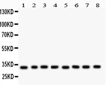
Anti-SMN1/2 Picoband antibody, Western blotting. All lanes: Anti SMN1/2 at 0.5 ug/ml. Lane 1: Rat Brain Tissue Lysate at 50 ug. Lane 2: Mouse Brain Tissue Lysate at 50 ug. Lane 3: Rat Liver Tissue Lysate at 50 ug. Lane 4: Mouse Liver Tissue Lysate at 50 ug. Lane 5: 293T Whole Cell Lysate at 40 ug. Lane 6: SMMC Whole Cell Lysate at 40 ug. Lane 7: HEPG2 Whole Cell Lysate at 40 ug. Lane 8: HELA Whole Cell Lysate at 40 ug. Predicted bind size: 32 KD. Observed bind size: 32 KD.
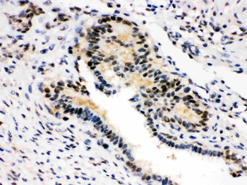
Anti-SMN1/2 Picoband antibody, IHC(P): Human Mammary Cancer Tissue.
Anti-SMN1/2 Picoband antibody, IHC(P): Mouse Brain Tissue.
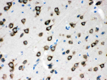
Anti-SMN1/2 Picoband antibody, IHC(P): Rat Brain Tissue.
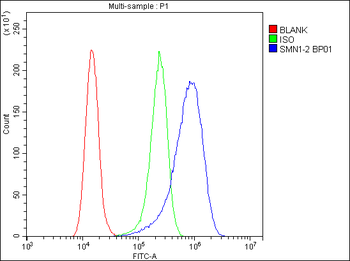
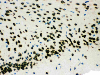
Flow Cytometry analysis of A431 cells using anti-SMN1/2 antibody. Overlay histogram showing A431 cells (Blue line). To facilitate intracellular staining, cells were fixed with 4% paraformaldehyde and permeabilized with permeabilization buffer. The cells were blocked with 10% normal goat serum. And then incubated with rabbit anti-SMN1/2 Antibody (1 µg/1x10^6 cells) for 30 min at 20°C. DyLight®488 conjugated goat anti-rabbit IgG (5-10 µg/1x10^6 cells) was used as secondary antibody for 30 minutes at 20°C. Isotype control antibody (Green line) was rabbit IgG (1 µg/1x10^6) used under the same conditions. Unlabelled sample without incubation with primary antibody and secondary antibody (Red line) was used as a blank control.
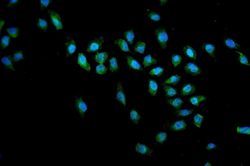
IF analysis of SMN1/2 using anti-SMN1/2 antibody. SMN1/2 was detected in immunocytochemical section of U20S cells. Enzyme antigen retrieval was performed using IHC enzyme antigen retrieval reagent for 15 mins. The cells were blocked with 10% goat serum. And then incubated with 2 µg/mL rabbit anti-SMN1/2 Antibody overnight at 4°C. DyLight®488 Conjugated Goat Anti-Rabbit IgG was used as secondary antibody at 1:100 dilution and incubated for 30 minutes at 37°C. The section was counterstained with DAPI. Visualize using a fluorescence microscope and filter sets appropriate for the label used.
Quick Database Links
Gene Symbol
Survival motor neuron protein
UniProt
UniProt Details
− No UniProt data available
Documents Download
Datasheet
Product Information
Request a Document
Protocol Information
WB
Western Blot (IB, immunoblot)
IHC
Immunohistochemistry
FC
Flow Cytometry
IF
Immunofluorescence
ICC
Immunocytochemistry
SMN1/2 Rabbit Polyclonal Antibody (orb251581)

- 0.0
Based on 0 reviews
Participating in our Biorbyt product reviews program enables you to support fellow scientists by sharing your firsthand experience with our products.
Login to Submit a ReviewAvailable Sizes
Select a size below
Free Secondary Antibody (20 ul)0/0
Please add an antibody product to your cart first.