You have no items in your shopping cart.
Description
Research Area
Cell Biology
Images & Validation
−Item 1 of 8
| Tested Applications | ELISA, IF, IHC, WB |
|---|---|
| Dilution Range | ELISA: 1:10,000 - 1:50,000, IHC: 1:50-1:200, IF: 1:50-1:200, WB: 1:1000-1:5000 |
| Reactivity | Human, Mouse |
| Application Notes |
Key Properties
−| Antibody Type | Primary Antibody |
|---|---|
| Host | Rabbit |
| Clonality | Polyclonal |
| Isotype | IgG |
| Immunogen | This antibody was affinity purified from whole rabbit serum prepared by repeated immunizations with a synthetic peptide corresponding to an extracellular region of human beta amyloid conjugated to KLH using maleimide. |
| Target | APP |
| Purity | This affinity purified antibody is directed against extracellular region of beta amyloid and is useful in determining its presence in various assays. Polyclonal anti-beta amyloid detects human and mouse beta amyloid. Blast analysis of the sequence of the immunogen shows 100% identity with Human, Guinea Pig, Pig, Cyno Monkey, Dog, Polar Bear, Rabbit, Chimp, Squirrel monkey, and Sheep. Cross reactivity with beta amyloid from other species is likely but has not been determined. |
| Conjugation | Unconjugated |
Storage & Handling
−| Storage | Store vial at -20° C or below prior to opening. This vial contains a relatively low volume of reagent (25 µL). To minimize loss of volume dilute 1:10 by adding 225 µL of the buffer stated above directly to the vial. Recap, mix thoroughly and briefly centrifuge to collect the volume at the bottom of the vial. Use this intermediate dilution when calculating final dilutions as recommended below. Store the vial at -20°C or below after dilution. Avoid cycles of freezing and thawing. |
|---|---|
| Form/Appearance | Liquid (sterile filtered) |
| Buffer/Preservatives | Preservative: 0.01% (w/v) Sodium Azide. Stabilizer: None; Buffer: 0.02 M Potassium Phosphate, 0.15 M Sodium Chloride, pH 7.2 |
| Concentration | 0.93 mg/mL |
| Expiration Date | 12 months from date of receipt. |
| Dry Ice Shipping | Please note: This product requires shipment on dry ice. A dry ice surcharge will apply. |
| Disclaimer | For research use only |
Alternative Names
−rabbit anti-Beta Amyloid Antibody, ß-amyloid, Amyloid beta A4 protein, Alzheimer disease amyloid protein, Beta amyloid, A-beta, ABPP, APPI, Beta-amyloid precursor protein
Similar Products
−- Item 1 of 8
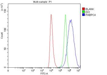
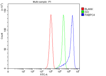
APP-1/PABPC4/APP Rabbit Polyclonal Antibody [orb1173479]
FC, ICC, IF, IHC, WB
Human, Mouse, Rat
Rabbit
Polyclonal
Unconjugated
100 μg - Item 1 of 8
- Item 1 of 7
beta Amyloid/APP Rabbit Polyclonal Antibody [orb196261]
ICC, IF, IHC, WB
Human, Mouse, Rat
Rabbit
Polyclonal
Unconjugated
100 μg - Item 1 of 7
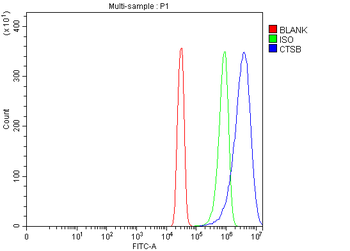
Cathepsin B/CTSB Rabbit Polyclonal Antibody [orb1098012]
ELISA, FC, IHC, WB
Human, Mouse, Rat
Rabbit
Polyclonal
Unconjugated
100 μg - Item 1 of 6


Quality Guarantee
Explore bioreagents carefree to elevate your research. All our products are rigorously tested for performance. If a product does not perform as described on its datasheet, our scientific support team will provide expert troubleshooting, a prompt replacement, or a refund. For full details, please see our Terms & Conditions and Buying Guide. Contact us at [email protected].
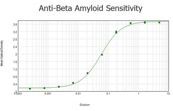
ELISA results of purified Rabbit anti-Beta Amyloid Antibody tested against BSA-conjugated peptide of immunizing peptide. Each well was coated in duplicate with 0.1 µg of conjugate. The starting dilution of antibody was 5 µg/ml and the X-axis represents the Log10 of a 3-fold dilution. This titration is a 4-parameter curve fit where the IC50 is defined as the titer of the antibody. Assay performed using 3% fish gel, Goat anti-Rabbit IgG Antibody Peroxidase Conjugated (Min X Bv Ch Gt GP Ham Hs Hu Ms Rt & Sh Serum Proteins) (p/n orb347654) and TMB ELISA Peroxidase Substrate (p/n orb348651).

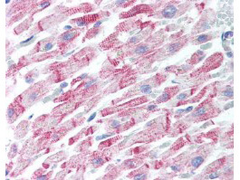
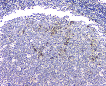
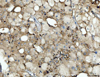
Human Heart (formalin-fixed, paraffin-embedded) stained with Anti-Beta Amyloid Antibody at 5 ug/ml followed by biotinylated goat anti-rabbit IgG secondary antibody, alkaline phosphatase-streptavidin and chromogen.
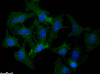
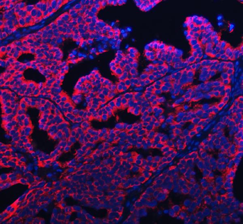
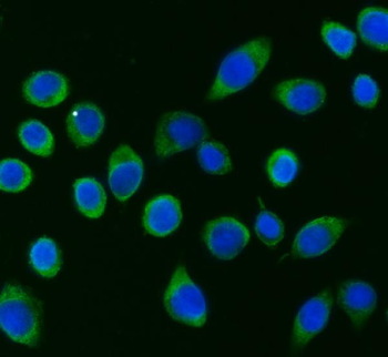
Immunofluorescence microscopy of Rabbit Anti-Beta Amyloid antibody using HeLa cells fixed with MeOH. Anti-Beta Amyloid Antibody was used at 1 µg/ml, O/N at 4°C. Secondary antibody: Anti-RABBIT IgG DyLight™ 488 Conjugated Preadsorbed at 2 ug/ml for 1 h at RT. Localization: APP is a cell surface protein that rapidly becomes internalized to endosomes and lysosomes. Some APP accumulates in secretory transport vesicles. Colocalizes with other proteins in a vesicular pattern in cytoplasm and perinuclear regions. Staining: Amyloid beta as green fluorescent signal with DAPI (blue) nuclear counterstain.
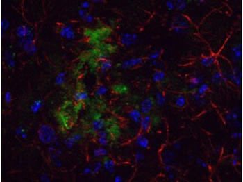
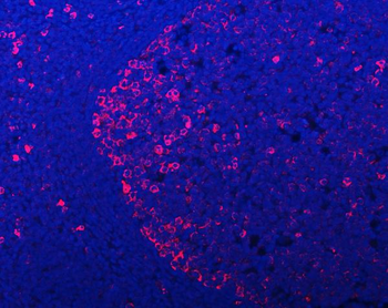
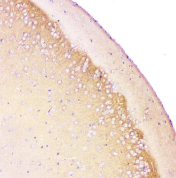
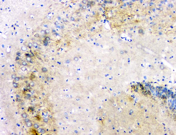
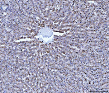
Immunohistochemical detection of beta Amyloid using Anti-Beta Amyloid Antibody on TG APP23 mouse brain cortex frozen sections. Anti-Beta Amyloid Antibody used at 1:200 and incubated for 2 hours in TBS/BSA with Tween and azide. Fluorescent labelled anti rabbit IgG was then added.
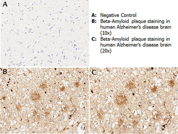
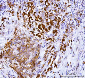
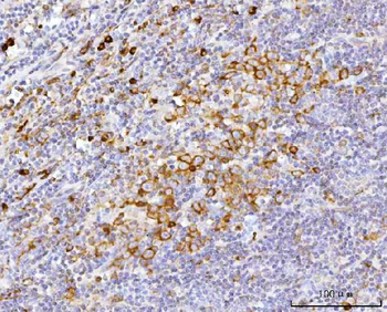
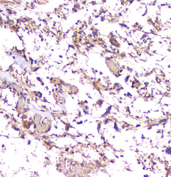
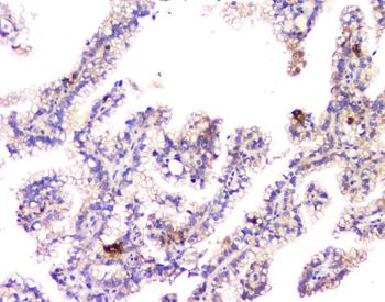
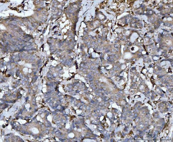
Immunohistochemistry with anti-beta amyloid antibody showing amyloid beta plaque staining in human Alzheimer's disease brain at 10x and 20x (B & C). Staining was performed on Leica Bond system using the standard protocol. Formalin fixed/paraffin embedded tissue sections were subjected to antigen retrieval with E1 (Leica Microsystems) retrieval solution for 20 min and then incubated with rabbit anti-beta amyloid antibody orb345371 at 1:100 dilution for 60 minutes. Biotinylated Anti-rabbit secondary antibody was used at 1:200 dilution to detect primary antibody. The reaction was developed using streptavidin-HRP conjugated compact polymer system and visualized with chromogen substrate, 3'3-diamino-benzidine substrate (DAB). The sections were then counterstained with hematoxylin to detect cell nuclei.

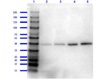
Western Blot of Rabbit Anti-Beta Amyloid Antibody. Lane 1: Opal Prestained Molecular Weight Marker. Lane 2: HEK293T Whole Cell Lysate. Lane 3: Mouse Brain Whole Cell Lysate. Lane 4: A-172 Whole Cell Lysate (p/n orb348708). Lane 5: Daudi Whole Cell Lysate (p/n orb692723). Load: 10 µg/lane. Primary Antibody: Anti-Beta Amyloid at 1:1000 overnight at 2-8°C. Secondary Antibody: Goat Anti-Rabbit IgG HRP Conjugated (p/n orb347654) at 1:70000 for 30 min at RT. Block: BlockOut Buffer (p/n orb348644). Predicted MW: ~40-50kDa. Observed MW: ~48kDa.
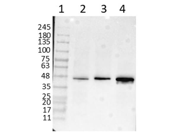
Western Blot of Rabbit Anti-Beta Amyloid Antibody. Lane 1: Opal Prestained Molecular Weight Marker. Lane 2: HEK293T Whole Cell Lysate. Lane 3: Mouse Brain Whole Cell Lysate. Lane 4: A-172 Whole Cell Lysate (p/n orb348708). Load: 10 µg/lane. Primary Antibody: Anti-Beta Amyloid at 1 µg/ml overnight at 2-8°C. Secondary Antibody: Goat Anti-Rabbit IgG HRP Conjugated (p/n orb347654) at 1:70000 for 30 min at RT. Block: BlockOut Buffer (p/n orb348644). Predicted MW: ~40-50kDa. Observed MW: ~48kDa.
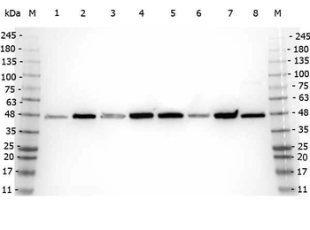
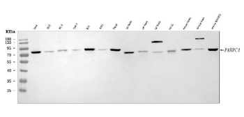
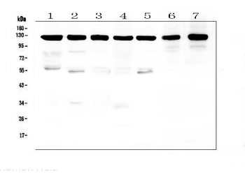
Western Blot of Rabbit anti-Beta Amyloid antibody. Marker: Opal Pre-stained ladder. Lane 1: HEK293 lysate (p/n orb348669). Lane 2: HeLa Lysate (p/n orb348668). Lane 3: MCF-7 Lysate (p/n orb348664). Lane 4: Jurkat Lysate. Lane 5: A431 Lysate (p/n orb348665). Lane 6: LNCaP Lysate (p/n orb348694). Lane 7: A-172 Lysate (p/n orb348708). Lane 8: NIH/3T3 Lysate (p/n orb348714). Load: 35 µg per lane. Primary antibody: Beta Amyloid antibody at 1:5000 for overnight at 4°C. Secondary antibody: Peroxidase rabbit secondary antibody at 1:30000 for 60 min at RT. Blocking Buffer: 1% Casein-TTBS for 30 min at RT. Predicted MW: ~40-50 kDa. Observed size: ~48 kDa for Beta Amyloid.
Quick Database Links
UniProt Details
− No UniProt data available
NCBI Reference Sequences
−Associated Accession Numbers
Curated reference sequences for the gene transcript and protein product| Protein | NP_000475.1 |
|---|
Documents Download
Datasheet
Product Information
Request a Document
Protocol Information
WB
Western Blot (IB, immunoblot)
IHC
Immunohistochemistry
IF
Immunofluorescence
ELISA
Enzyme-linked Immunosorbent Assay (EIA)
APP Antibody (orb345372)

- 0.0
Based on 0 reviews
Participating in our Biorbyt product reviews program enables you to support fellow scientists by sharing your firsthand experience with our products.
Login to Submit a ReviewAvailable Sizes
Select a size below
Free Secondary Antibody (20 ul)0/0
Please add an antibody product to your cart first.