You have no items in your shopping cart.
Featured
KO/KD
Validated
Validated
Description
Research Area
Cell Biology
Images & Validation
−Item 1 of 11
| Tested Applications | ELISA, ICC, IF, KO/KD Validated, WB |
|---|---|
| Reactivity | Human, Mouse |
| Predicted Reactivity | Rat |
Key Properties
−| Antibody Type | Primary Antibody |
|---|---|
| Host | Rabbit |
| Clonality | Polyclonal |
| Isotype | IgG |
| Immunogen | Anti-BIM antibody (orb1239258) was raised against a peptide corresponding to amino acids near the center of human BIM. The immunogen is located within amino acids 80-130 of BIM. |
| Target | BCL2L11 |
| Molecular Weight | Predicted: 22kDObserved: 22kD |
| Purification | Bim Antibody is affinity chromatography purified via peptide column. |
| Conjugation | Unconjugated |
Storage & Handling
−| Storage | Maintain refrigerated at 2-8°C for up to 2 weeks. For long term storage store at -20°C in small aliquots to prevent freeze-thaw cycles. |
|---|---|
| Form/Appearance | Liquid |
| Buffer/Preservatives | Bim Antibody is supplied in PBS containing 0.02% sodium azide. |
| Concentration | 1 mg/mL |
| Expiration Date | 12 months from date of receipt. |
| Disclaimer | For research use only |
Alternative Names
−Bim Antibody: BAM, BIM, BOD, Bcl-2-like protein 11, Bcl2-interacting mediator of cell death, Bcl2-L-11
Similar Products
−- Item 1 of 13
BCL2L11 Antibody [orb1239259]
ELISA, IF, IHC-P, KO/KD Validated, WB
Human, Mouse, Rat
Rabbit
Polyclonal
Unconjugated
0.1 mg, 0.02 mg - Item 1 of 4
BCL2L11 Antibody (Center) [orb1928438]
FC, IHC-P, WB
Mouse, Rat
Human
Rabbit
Polyclonal
Unconjugated
50 μl, 100 μl - Item 1 of 5
Bim/BCL2L11 Rabbit Polyclonal Antibody [orb1098118]
FC, IHC, WB
Human, Mouse
Rabbit
Polyclonal
Unconjugated
100 μg - Item 1 of 1
- Item 1 of 3


Quality Guarantee
Explore bioreagents carefree to elevate your research. All our products are rigorously tested for performance. If a product does not perform as described on its datasheet, our scientific support team will provide expert troubleshooting, a prompt replacement, or a refund. For full details, please see our Terms & Conditions and Buying Guide. Contact us at [email protected].

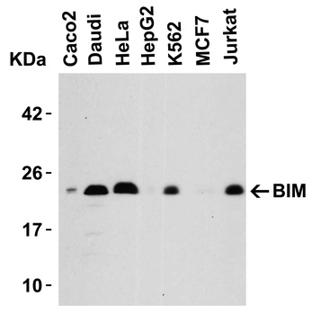
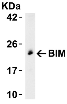
Western Blot Validation in Human Cell Lines. Loading: 15 µg of lysates per lane. Antibodies: BIM orb1239258, (5 µg/mL), 1h incubation at RT in 5% NFDM/TBST. Secondary: Goat anti-rabbit IgG HRP conjugate at 1:10000 dilution.

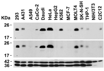
Independent Antibody Validation (IAV) via Protein Expression Profile in Cell Lines. Loading: 15 µg of lysates per lane. Antibodies: BIM orb1239259, (0.5 µg/mL), BIM orb1239258, (5 µg/mL), beta-actin (1 µg/mL) and GAPDH (0.02 µg/mL), 1h incubation at RT in 5% NFDM/TBST. Secondary: Goat anti-rabbit IgG HRP conjugate at 1:10000 dilution.
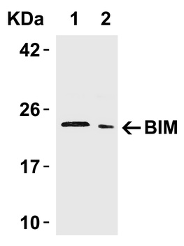
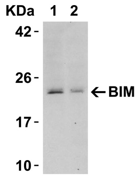
Western Blot Validation in Human Tissue. Loading: 15 µg of lysates per lane. Antibodies: BIM orb1239258, (5 µg/mL), 1h incubation at RT in 5% NFDM/TBST. Secondary: Goat anti-rabbit IgG HRP conjugate at 1:10000 dilution. Lane 1: Human urinary bladder, Lane 2: Human pancreas.
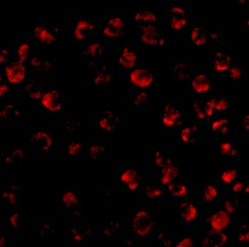
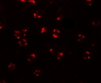
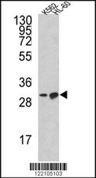
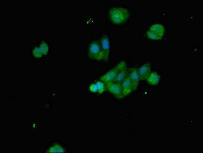
Immunofluorescence Validation of BIM in K562 Cells. Immunofluorescent analysis of 4% paraformaldehyde-fixed K562 cells labeling BIM with orb1239258 at 20 µg/mL, followed by goat anti-rabbit IgG secondary antibody at 1/500 dilution (red).
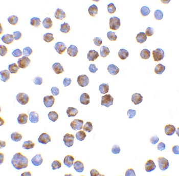
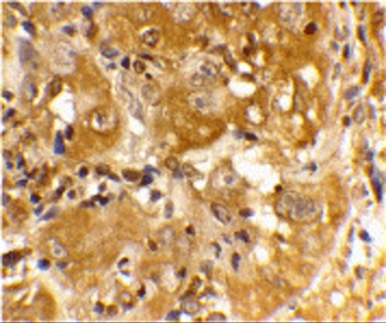
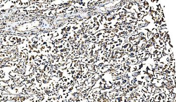
Immunocytochemistry Validation of BIM K562 Cells. Immunohistochemical analysis of K562 cells using anti-BIM antibody (orb1239258) at 10 µg/ml. Cells was fixed with formaldehyde and blocked with 10% serum for 1 h at RT; antigen retrieval was by heat mediation with a citrate buffer (pH6). Samples were incubated with primary antibody overnight at 4°C. A goat anti-rabbit IgG H&L (HRP) at 1/250 was used as secondary. Counter stained with Hematoxylin.
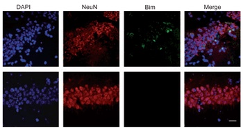
Induced Expression Validation of BIM in Mouse Hippocampus (Tsuchiya et al., 2011). The induction of Bim protein was detected by immunohistochemical analysis of mice after i.h. injection of epoxomicin with anti-BIM antibodies. Sections from epoxomicin-treated animals exhibited cells staining positive for Bim expression within the NeuN-positive population of neurons in the CA1 of the ipsilateral side. In contrast, Bim-positive cells were absent within the NeuNpositiveCA1 neurons on the contralateral side.

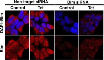
KD Validation of BIM in 293 Cells (Han et al., 2010). Immunofluorescence analysis with anti-BIM antibodies was performed for BIM in 293 cells transfected with control siRNA or BIM siRNA. BIM expression was disrupted after BIM siRNA knockdown.

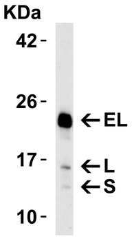
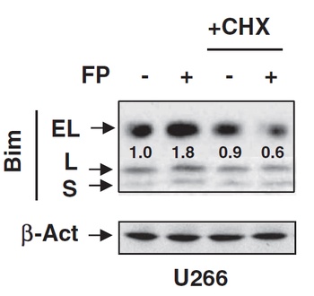
Regulated Expression Validation of BIM in U266 Cells (Chen et al., 2012). Immunoblot analysis was carried out to monitor protein expression of 3 isoforms (EL, L, and S) of Bim with anti-BIM antibodies. BIM expression was up-regulated by flavopiridol treatment, which was blocked by Cycloheximide in U266 cells.

WB KD Validation of BIM in 293 Cells (Han et al., 2010). Western blot analysis with anti-BIM antibodies was performed for BIM in 293 cells transfected with control siRNA or BIM siRNA. BIM expression was disrupted after BIM siRNA knockdown.

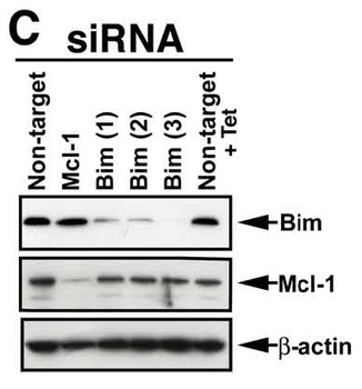
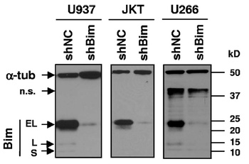
KD Validation of BIM in Human Cell Lines (Chen et al., 2009). Human leukemia (U937 and Jurkat) and myeloma (U266) cells were stably transfected with constructs encoding shBim or a scrambled sequence (shNC). Immunoblotting was preformed to monitor expression of Bim in these cells with anti-BIM antibodies. BIM expression was disrupted after shBIM.
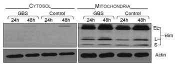
Localization Validation of BIM in Mouse Macrophages (Ulett et al., 2005). Immunoblots of subcellular fractions enriched for mitochondria and cytosol were used to determine BIM protein levels with anti-BIM antibodies in J774A cells. BIM is exclusively expressed in mitochondria.
Documents Download
Datasheet
Product Information
Request a Document
Protocol Information
WB
Western Blot (IB, immunoblot)
IF
Immunofluorescence
ICC
Immunocytochemistry
ELISA
Enzyme-linked Immunosorbent Assay (EIA)
BCL2L11 Antibody (orb1239258)

- 0.0
Based on 0 reviews
Participating in our Biorbyt product reviews program enables you to support fellow scientists by sharing your firsthand experience with our products.
Login to Submit a ReviewAvailable Sizes
Select a size below
Free Secondary Antibody (20 ul)0/0
Please add an antibody product to your cart first.