You have no items in your shopping cart.
Featured
Description
Research Area
Cell Biology
Images & Validation
−Item 1 of 11
| Tested Applications | FC, ICC, IF, IHC-Fr, IHC-P, WB |
|---|---|
| Dilution Range | WB=1:500-2000, IHC-P=1:100-500, IHC-F=1:100-500, ICC/IF=1:100-500, IF=1:100-500, Flow-Cyt=1μg/test |
| Reactivity | Human, Mouse, Rat |
| Predicted Reactivity | Bovine, Canine |
Related Conjugates & Formulations
−Key Properties
−| Antibody Type | Primary Antibody |
|---|---|
| Host | Rabbit |
| Clonality | Polyclonal |
| Isotype | IgG |
| Immunogen | KLH conjugated synthetic peptide derived from human Caspase-9 subunit p35 (11-120/416aa) |
| Target | CASP9 |
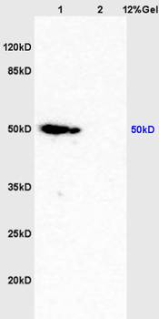
| Molecular Weight | 52 kDa |
| Purification | Affinity purified by Protein A |
| Conjugation | Unconjugated |
Storage & Handling
−| Storage | Maintain refrigerated at 2-8°C for up to 2 weeks. For long term storage store at -20°C in small aliquots to prevent freeze-thaw cycles. |
|---|---|
| Form/Appearance | Liquid |
| Buffer/Preservatives | 0.01M TBS (pH7.4) with 1% rAlbumin, 0.02% Proclin300 and 50% Glycerol. |
| Concentration | 1mg/ml |
| Expiration Date | 12 months from date of receipt. |
| Disclaimer | For research use only |
Alternative Names
−APAF-3; APAF3; ICE-LAP6; MCH6; PPP1R56; CASP-9; Caspase-9; Casp-9-CTD; Casp9_v1; CASP9_HUMAN; CASP9; Apoptotic protease Mch-6; Apoptotic protease-activating factor 3 (APAF-3); ICE-like apoptotic protease 6 (ICE-LAP6); 3.4.22.62; Q4FJK5_MOUSE; Apoptotic protease-activating factor 3; ICE-like apoptotic protease 6; Q920G4_RAT;
Similar Products
−- Item 1 of 9
Caspase-9 Rabbit Polyclonal Antibody [orb500803]
FC, ICC, IF, IHC-Fr, IHC-P, WB
Bovine, Porcine, Rabbit, Sheep
Human, Mouse, Rat
Rabbit
Polyclonal
Unconjugated
50 μl, 100 μl, 200 μl - Item 1 of 7
Caspase-9 Rabbit Polyclonal Antibody [orb10243]
FC, ICC, IF, IHC-Fr, IHC-P, WB
Bovine, Porcine, Rabbit, Sheep
Human, Mouse, Rat
Rabbit
Polyclonal
Unconjugated
50 μl, 100 μl, 200 μl - Item 1 of 7
Phospho-Caspase-9 (Thr125) Rabbit Polyclonal Antibody [orb5767]
FC, IF, IHC-Fr, IHC-P, WB
Mouse, Rat
Human, Mouse, Rat
Rabbit
Polyclonal
Unconjugated
50 μl, 100 μl, 200 μl - Item 1 of 7
Caspase-9 p10 Rabbit Polyclonal Antibody [orb1677]
FC, IF, IHC-Fr, IHC-P, WB
Bovine, Canine, Equine, Gallus, Porcine, Rabbit
Human, Mouse, Rat
Rabbit
Polyclonal
Unconjugated
100 μl, 200 μl, 50 μl - Item 1 of 6
Caspase-9/CASP9 Rabbit Polyclonal Antibody [orb763020]
ELISA, FC, IHC, WB
Human
Rabbit
Polyclonal
Unconjugated
100 μg


Quality Guarantee
Explore bioreagents carefree to elevate your research. All our products are rigorously tested for performance. If a product does not perform as described on its datasheet, our scientific support team will provide expert troubleshooting, a prompt replacement, or a refund. For full details, please see our Terms & Conditions and Buying Guide. Contact us at [email protected].
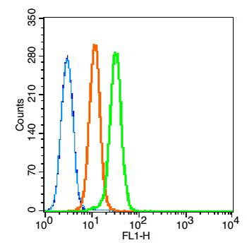
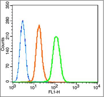
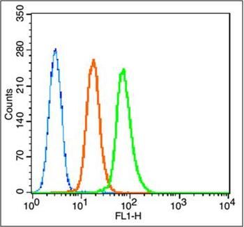
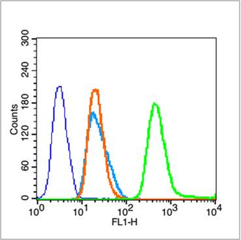
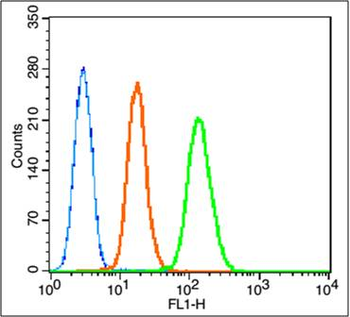
Blank control: K562 (blue). Primary Antibody: Rabbit Anti-caspase-9 antibody (orb10242, Green), Dilution: 1 µg in 100 µL 1X PBS containing 0.5% BSA, Isotype Control Antibody: Rabbit IgG (orange), used under the same conditions, Secondary Antibody: Goat anti-rabbit IgG-FITC (white blue), Dilution: 1:200 in 1 X PBS containing 0.5% BSA. Protocol, The cells were fixed with 80% methanol (5 min) and and then permeabilized with 0.01M PBS-Tween for 20 min. Primary antibody (orb10242, 1 µg/1x10^6 cells) were incubated for 30 min at room temperature, followed by 1 X PBS containing 0.5% BSA + 10% goat serum (30 min) to block non-specific protein-protein interactions. Then the Goat Anti-rabbit IgG/FITC antibody was added into the blocking buffer mentioned above to react with the primary antibody at 1/200 dilution for 30 min at room temperature. Acquisition of 20000 events was performed.
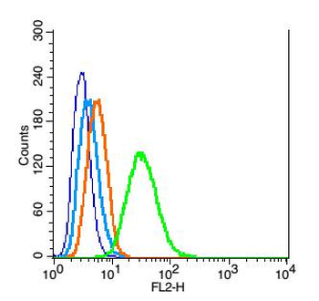
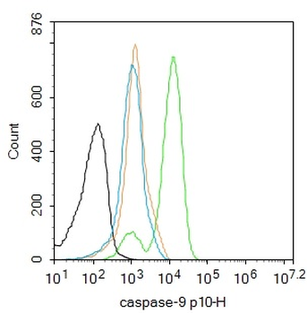
Blank control: RSC96 (blue), the cells were fixed with 2% paraformaldehyde (10 min) and then permeabilized with ice-cold 90% methanol for 30 min on ice. Isotype Control Antibody: Rabbit IgG (orange), Secondary Antibody: Goat anti-rabbit IgG-PE (white blue), Dilution: 1:200 in 1 X PBS containing 0.5% BSA, Primary Antibody Dilution: 1 µg in 100 µL 1X PBS containing 0.5% BSA (green).
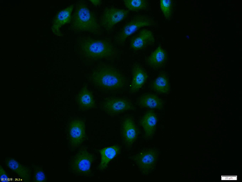
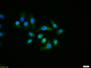
HepG2 cell, 4% Paraformaldehyde-fixed, Triton X-100 at room temperature for 20 min, Blocking buffer (normal goat serum) at 37°C for 20 min, Antibody incubation with (Caspase-9) polyclonal Antibody, Unconjugated (orb10242) 1:100, 90 minutes at 37°C, followed by a conjugated Goat Anti-Rabbit IgG antibody at 37°C for 90 minutes, DAPI (blue) was used to stain the cell nuclei.
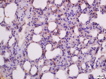
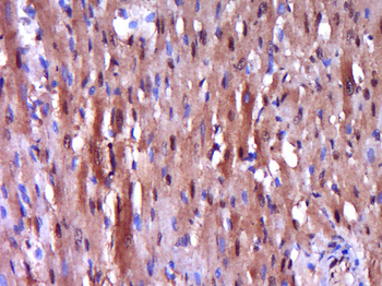
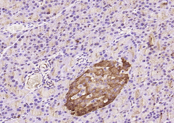
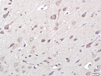
Paraformaldehyde-fixed, paraffin embedded (rat lung), Antigen retrieval by boiling in sodium citrate buffer (pH6.0) for 15 min, Block endogenous peroxidase by 3% hydrogen peroxide for 20 minutes, Blocking buffer (normal goat serum) at 37°C for 30 min, Antibody incubation with (Insulin like growth factor 1) Polyclonal Antibody, Unconjugated at 1:400 overnight at 4°C, followed by a conjugated secondary antibody for 20 minutes and DAB staining.
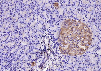
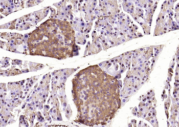
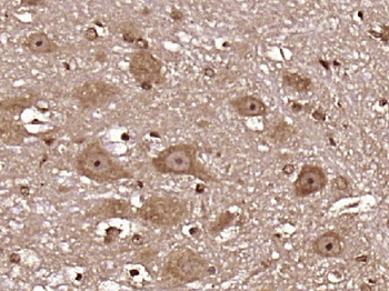
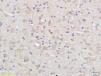
Paraformaldehyde-fixed, paraffin embedded (rat pancreas), Antigen retrieval by boiling in sodium citrate buffer (pH6.0) for 15 min, Block endogenous peroxidase by 3% hydrogen peroxide for 20 minutes, Blocking buffer (normal goat serum) at 37°C for 30 min, Antibody incubation with (Caspase-9) Polyclonal Antibody, Unconjugated (orb10242) at 1:200 overnight at 4°C, followed by operating according to SP Kit (Rabbit) instructionsand DAB staining.
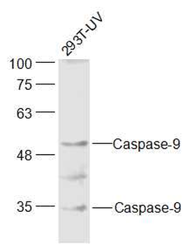
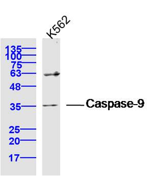
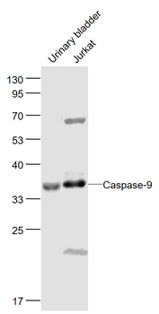
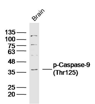
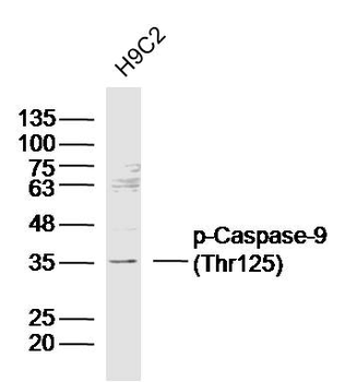
Sample: 293T-UV Cell (Human) Lysate at 30 ug, Primary: Anti-Caspase-9 at 1/300 dilution, Secondary: IRDye800CW Goat Anti-Rabbit IgG at 1/20000 dilution, Predicted band size: 35/50 kD, Observed band size: 35/50 kD.
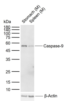
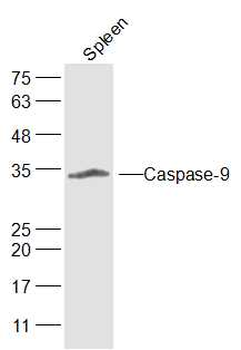
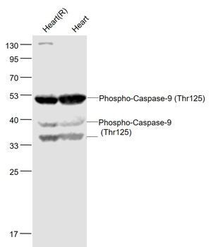
Sample: Lane 1: Mouse Stomach tissue lysates, Lane 2: Mouse Spleen tissue lysates, Primary: Anti-Caspase-9 (orb10242) at 1/1000 dilution, Secondary: IRDye800CW Goat Anti-Rabbit IgG at 1/20000 dilution, Predicted band size: 35/50 kDa, Observed band size: 52 kDa.
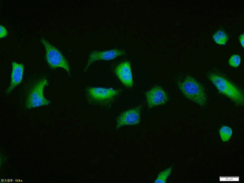
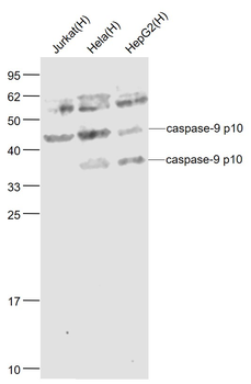
Tissue/cell: Hela cell, 4% Paraformaldehyde-fixed, Triton X-100 at room temperature for 20 min, Blocking buffer (normal goat serum) at 37°C for 20 min, Antibody incubation with (Caspase-9) polyclonal Antibody, Unconjugated (orb10242) 1:100, 90 minutes at 37°C, followed by a FITC conjugated Goat Anti-Rabbit IgG antibody at 37°C for 90 minutes, DAPI (blue) was used to stain the cell nuclei.
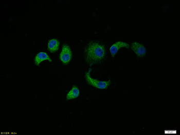
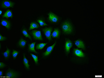
Tissue/cell: HepG2 cell, 4% Paraformaldehyde-fixed, Triton X-100 at room temperature for 20 min, Blocking buffer (normal goat serum) at 37°C for 20 min, Antibody incubation with (Caspase-9) polyclonal Antibody, Unconjugated (orb10242) 1:100, 90 minutes at 37°C, followed by a FITC conjugated Goat Anti-Rabbit IgG antibody at 37°C for 90 minutes, DAPI (blue) was used to stain the cell nuclei.
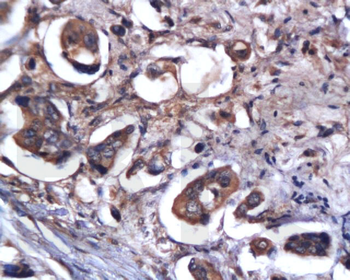
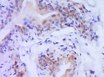
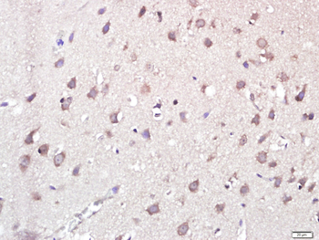
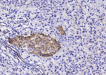
Tissue/cell: human colon carcinoma, 4% Paraformaldehyde-fixed and paraffin-embedded, Antigen retrieval: citrate buffer (0.01M, pH 6.0), Boiling bathing for 15 min, Block endogenous peroxidase by 3% Hydrogen peroxide for 30 min, Blocking buffer (normal goat serum) at 37℃ for 20 min, Incubation: Anti-Caspase-9 Polyclonal Antibody, Unconjugated (orb10242) 1:200, overnight at 4°C, followed by conjugation to the secondary antibody and DAB staining.
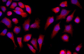
Tissue/cell: MCF-7 cell, 4% Paraformaldehyde-fixed, Triton X-100 at room temperature for 20 min, Blocking buffer (normal goat serum) at 37°C for 20 min, Antibody incubation with (Caspase-9) Polyclonal Antibody, Unconjugated (orb10242) 1:50, 90 minutes at 37°C, followed by a conjugated Goat Anti-Rabbit IgG antibody (orb868589) at 37°C for 90 minutes, DAPI (blue) was used to stain the cell nuclei.
Quick Database Links
Gene Symbol
CASP9
UniProt
UniProt Details
− No UniProt data available
Documents Download
Datasheet
Product Information
Request a Document
Protocol Information
WB
Western Blot (IB, immunoblot)
IHC-P
Immunohistochemistry Paraffin
IHC-Fr
Immunohistochemistry Frozen
FC
Flow Cytometry
IF
Immunofluorescence
ICC
Immunocytochemistry
Chen, Liwen et al. Combination of gemcitabine and erlotinib inhibits recurrent pancreatic cancer growth in mice via the JAK-STAT pathway Oncol Rep, 39, 1081-1089 (2018)
Caspase-9 Rabbit Polyclonal Antibody (orb10242)

- 0.0
Based on 0 reviews
Participating in our Biorbyt product reviews program enables you to support fellow scientists by sharing your firsthand experience with our products.
Login to Submit a ReviewAvailable Sizes
Select a size below
Free Secondary Antibody (20 ul)0/0
Please add an antibody product to your cart first.