You have no items in your shopping cart.
Featured
Description
Research Area
Cell Biology
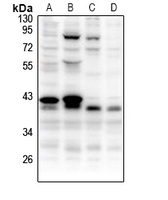
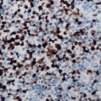
Images & Validation
−
Item 1 of 3
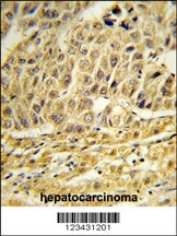
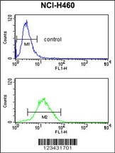
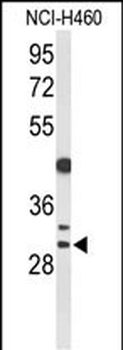
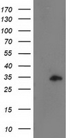
| Tested Applications | FC, IHC-P, WB |
|---|---|
| Reactivity | Human |
| Application Notes |
Key Properties
−| Antibody Type | Primary Antibody |
|---|---|
| Host | Rabbit |
| Clonality | Polyclonal |
| Isotype | Rabbit Ig |
| Immunogen | This CCNB1IP1 antibody is generated from rabbits immunized with a KLH conjugated synthetic peptide between 199-228 amino acids from the C-terminal region of human CCNB1IP1. |
| Target | CCNB1IP1 |
| Molecular Weight | 32 kDa |
| Purification | This antibody is purified through a protein A column, followed by peptide affinity purification. |
| Conjugation | Unconjugated |
Storage & Handling
−| Storage | Maintain refrigerated at 2-8°C for up to 2 weeks. For long term storage store at -20°C in small aliquots to prevent freeze-thaw cycles. |
|---|---|
| Form/Appearance | Liquid |
| Buffer/Preservatives | Supplied in PBS with 0.09% (W/V) sodium azide. |
| Concentration | batch dependent |
| Expiration Date | 12 months from date of receipt. |
| Disclaimer | For research use only |
Alternative Names
−E3 ubiquitin-protein ligase CCNB1IP1, 632-, Cyclin-B1-interacting protein 1, Human enhancer of invasion 10, CCNB1IP1, C14orf18, HEI10
Similar Products
−- Item 1 of 3
- Item 1 of 1
- Item 1 of 1
CCNB1IP1 Antibody [orb1323419]
FC, IHC, WB
Human, Mouse, Rat
Mouse
Monoclonal
Unconjugated
100 μl, 50 μl, 30 μl - Item 1 of 2
CCNB1IP1 Rabbit Polyclonal Antibody [orb319013]
IHC, WB
Human, Mouse, Rat
Rabbit
Polyclonal
Unconjugated
30 μl, 100 μl, 200 μl, 50 μl - Item 1 of 1


Quality Guarantee
Explore bioreagents carefree to elevate your research. All our products are rigorously tested for performance. If a product does not perform as described on its datasheet, our scientific support team will provide expert troubleshooting, a prompt replacement, or a refund. For full details, please see our Terms & Conditions and Buying Guide. Contact us at [email protected].
Protocol Information
WB
Western Blot (IB, immunoblot)
IHC-P
Immunohistochemistry Paraffin
FC
Flow Cytometry
Available Sizes
Select a size below
Free Secondary Antibody (20 ul)0/0
Please add an antibody product to your cart first.