You have no items in your shopping cart.
Description
Research Area
Cell Biology
Images & Validation
−Item 1 of 7
| Tested Applications | ICC, IF, IHC-P, WB |
|---|---|
| Dilution Range | WB - 1:1000, IHC-P - 1:100-500, ICC - 1:10-50 |
| Reactivity | Human, Mouse |
| Predicted Reactivity | Rat |
Key Properties
−| Host | Rabbit |
|---|---|
| Clonality | Polyclonal |
| Isotype | Rabbit IgG |
| Conjugation | Unconjugated |
Storage & Handling
−| Storage | Maintain refrigerated at 2-8°C for up to 2 weeks. For long term storage store at -20°C in small aliquots to prevent freeze-thaw cycles |
|---|---|
| Form/Appearance | Purified polyclonal antibody supplied in PBS with 0.09% (W/V) sodium azide. This antibody is purified through a protein A column, followed by peptide affinity purification. |
| Expiration Date | 12 months from date of receipt. |
| Disclaimer | For research use only |
Similar Products
−- Item 1 of 4
Cleaved LC3A Antibody [orb1787989]
IF, WB
Human
Rabbit
Polyclonal
Unconjugated
- Item 1 of 2
Cleaved LC3 Rabbit Polyclonal Antibody [orb628392]
ELISA, IHC, WB
Mouse, Rat
Rabbit
Polyclonal
Unconjugated
50 μg, 100 μg 


Quality Guarantee
Explore bioreagents carefree to elevate your research. All our products are rigorously tested for performance. If a product does not perform as described on its datasheet, our scientific support team will provide expert troubleshooting, a prompt replacement, or a refund. For full details, please see our Terms & Conditions and Buying Guide. Contact us at [email protected].
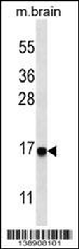
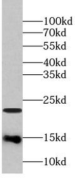
Western blot analysis of anti-cleaved-LC3 (APG8a) Pab in mouse brain tissue lysate. Cleaved-LC3 (APG8a) was detected using the purified Pab.
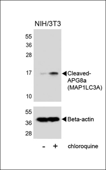
Western blot analysis of lysates from NIH/3T3 cells, untreated or treated with chloroquine, using Cleaved-APG8a (MAP1LC3A) (upper) or Beta-actin (lower).
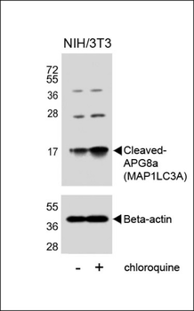
Western blot analysis of lysates from NIH/3T3 cells, untreated or treated with chloroquine, using Cleaved-APG8a (MAP1LC3A) (upper) or Beta-actin (lower).
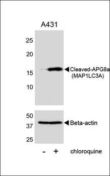
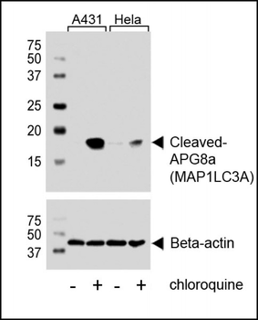
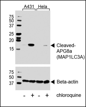
Western blot analysis of lysates from A431 cell line, untreated or treated with chloroquine, 100ng/ml, using Cleaved-APG8a (MAP1LC3A) (upper) or Beta-actin (lower).
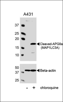
Western blot analysis of lysates from A431 cell line, untreated or treated with chloroquine, 100ng/ml, using Cleaved-APG8a (MAP1LC3A) Antibody (upper) or Beta-actin (lower).
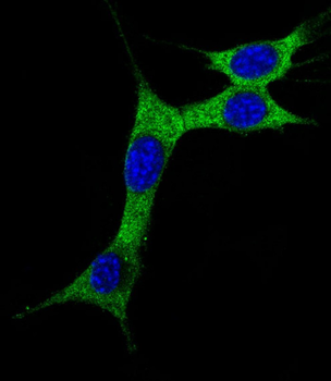
Immunofluorescent analysis of 4% paraformaldehyde-fixed, 0.1% Triton X-100 permeabilized NIH/3T3 (Mouse mouse embryonic fibroblasts cell line) cells labeling MAP1LC3A at 1/25 dilution, followed by Alexa Fluor 488-conjugated goat anti-rabbit IgG secondary antibody at 1/400 dilution (green). The nuclear counter stain is DAPI (blue). Immunofluorescence image showing cytoplasm on NIH/3T3 cell line.
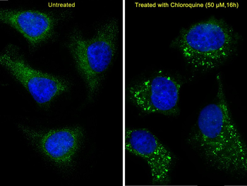
Immunofluorescent analysis of 4% paraformaldehyde-fixed, 0.1% Triton X-100 permeabilized Hela (Human Cervical epithelial adenocarcinoma cell line) cells labeling MAP1LC3A at 1/25 dilution, followed by Alexa Fluor 488-conjugated goat anti-rabbit IgG secondary antibody at 1/400 dilution (green). Immunofluorescence image showing vesicles staining on Hela cell line.The nuclear counter stain is DAPI (blue). The right image is Hela cells treated with Chloroquine 50μM for 16 h.
Documents Download
Datasheet
Product Information
Request a Document
Protocol Information
WB
Western Blot (IB, immunoblot)
IHC-P
Immunohistochemistry Paraffin
IF
Immunofluorescence
ICC
Immunocytochemistry
Filter by Applications
Filter by Species
Gopinath Mukherjee et al. TREATMENT OF MICE WITH TLR4 AND IFN-γ ANTIBODY AND EXOGENOUS IL-10 MODULATES BCL2 AND LC3 EXPRESSION IN RANKL/M-CSF PLUS LPS STIMULATED BONE MARROW MACROPHAGES-LEADING TO BONE RESORPTION researchgate.net, (2025)
Applications
WB
Reactivity
Mouse
Cleaved LC3A Antibody (orb1933527)

- 0.0
Based on 0 reviews
Participating in our Biorbyt product reviews program enables you to support fellow scientists by sharing your firsthand experience with our products.
Login to Submit a ReviewAvailable Sizes
Select a size below