You have no items in your shopping cart.
Featured
Description
Research Area
Epigenetics & Chromatin
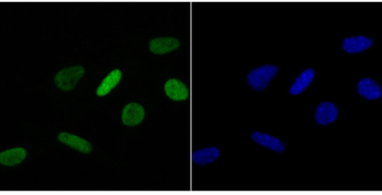
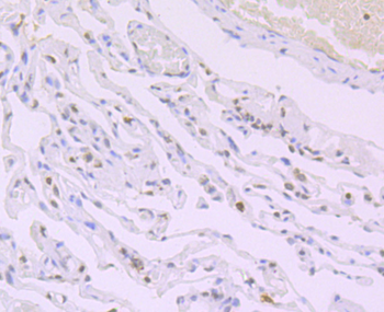
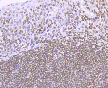
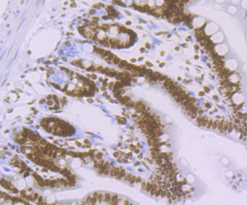
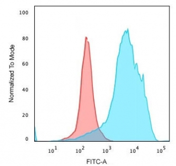
Images & Validation
−
Item 1 of 9
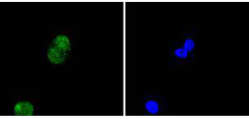
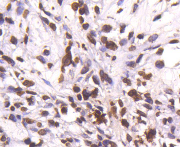
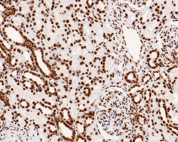
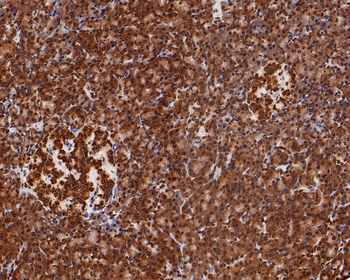
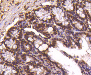
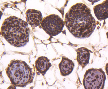
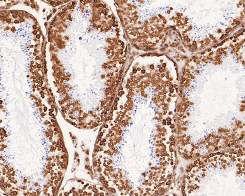
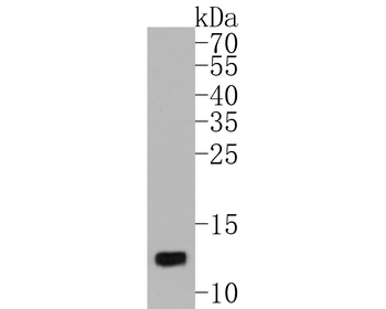
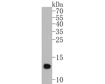
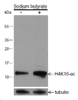
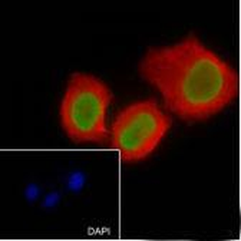
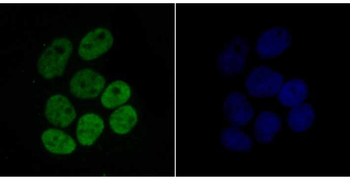
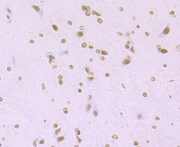
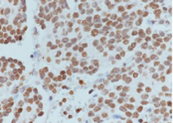
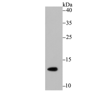
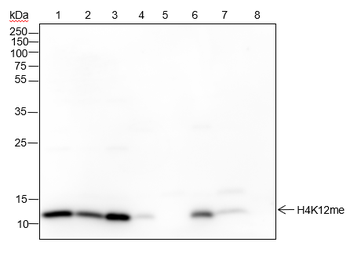
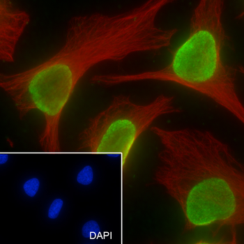
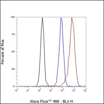
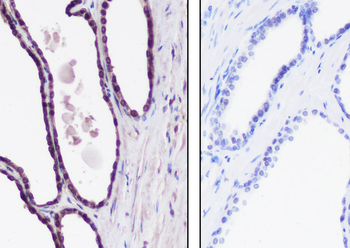
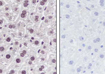
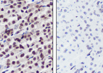
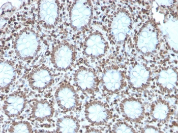
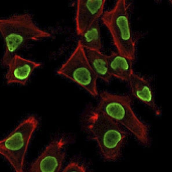
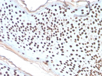
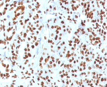
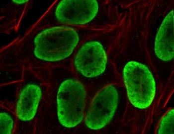
| Tested Applications | FC, IF, IHC-Fr, IHC-P, WB |
|---|---|
| Dilution Range | WB=1:500-2000, IHC-P=1:200-1000, IHC-F=1:200-1000, IF=1:200-1000, Flow-Cyt=1ug/Test |
| Reactivity | Human, Mouse, Rat |
| Predicted Reactivity | Mouse, Rat |
Key Properties
−| Antibody Type | Primary Antibody |
|---|---|
| Host | Rabbit |
| Clonality | Recombinant |
| Isotype | IgG |
| Clone No. | B4I4 |
| Immunogen | A synthesized peptide derived from human Histone H4 (80-103/103aa) |
| Target | H4C1 |
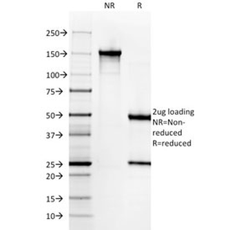
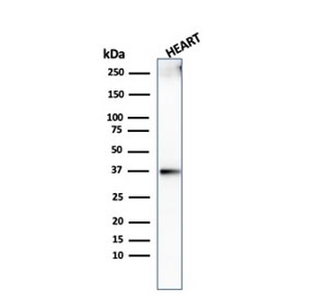
| Molecular Weight | 11 kDa |
| Purification | Affinity purified by Protein A |
| Conjugation | Unconjugated |
Storage & Handling
−| Storage | Maintain refrigerated at 2-8°C for up to 2 weeks. For long term storage store at -20°C in small aliquots to prevent freeze-thaw cycles. |
|---|---|
| Form/Appearance | Liquid |
| Buffer/Preservatives | 0.01M TBS (pH7.4) with 1% rAlbumin, 0.02% Proclin300 and 50% Glycerol. |
| Concentration | 1mg/ml |
| Expiration Date | 12 months from date of receipt. |
| Disclaimer | For research use only |
Alternative Names
−H4-16; H4/p; H4C1; H4C11; H4C12; H4C13; H4C14; H4C15; H4C2; H4C3; H4C4; H4C5; H4C6; H4C8; H4C9; HIST4H4; H4/o; H4C16; HIST2H4B; H4/m; H4FM; H4M; HIST1H4I; TEBIVANED4; TEVANED4; H4FA; HIST1H4A; H4/b; H4FB; HIST1H4D; dJ221C16.9; H4; H4/c; H4FC; HIST1H4F; H4/d; H4F2iii; H4FD; HIST1H4K; dJ160A22.1; H4/e; H4F2iv; H4FE; HIST1H4J; TEBIVANED2; TEVANED2; dJ160A22.2; H4/g; H4FG; HIST1H4C; TEBIVANED1; TEVANED1; dJ221C16.1; H4/h; H4FH; HIST1H4H; H4/I; H4FI; HIST1H4B; H4/j; H4FJ; HIST1H4E; TEBIVANED3; TEVANED3; H4.k; H4/k; H4FK; HIST1H4L; FO108; H4/n; H4F2; H4FN; HIST2H4; HIST2H4A; Gm11275; H4f16; Hist1h4m; Hist1h4n; B130044J01Rik; 1700024H08Rik; H2c8; H4_DROME; His4; His4r; His4:CG31611; His4:CG33869; His4:CG33871; His4:CG33873; His4:CG33875; His4:CG33877; His4:CG33879; His4:CG33881; His4:CG33883; His4:CG33885; His4:CG33887; His4:CG33889; His4:CG33891; His4:CG33893; His4:CG33895; His4:CG33897; His4:CG33899; His4:CG33901; His4:CG33903; His4:CG33905; His4:CG33907; His4:CG33909; H4r; H4_HUMAN; H4/A; H4FO; H4_MOUSE; H4-53; H4-12; H4_SCHPO; hhf1; hhf2; hhf3; h4.1; h4.2; h4.3; H4 clustered histone 1; H4 histone family, member A; histone 1, H4a; histone cluster 1, H4a; histone cluster 1 H4 family member a
Similar Products
−- Item 1 of 10
Histone H4 (acetyl K16) Recombinant Rabbit Monoclonal Antibody [orb1499354]
ChIP, FC, ICC, IF, IHC-Fr, IHC-P, WB
Human, Mouse, Rat
Human, Mouse, Rat
Rabbit
Recombinant
Unconjugated
50 μl, 100 μl - Item 1 of 6
Mono-Methyl-Histone H4 (Lys12) Recombinant Rabbit Monoclonal Antibody [orb1172948]
ChIP, FC, ICC, IF, IHC-Fr, IHC-P, IP, WB
Human, Mouse, Rat
Human, Mouse, Rat
Rabbit
Recombinant
Unconjugated
50 μl, 100 μl, 25 μl - Item 1 of 6
Recombinant Histone H1 Antibody [orb639702]
FACS, IF, IHC-P, WB
Human, Mouse, Rat
Rabbit
Recombinant
Unconjugated
100 μg, 20 μg - Item 1 of 6
Recombinant Histone H1 Antibody [orb2643346]
FACS, IF, IHC-P, WB
Human, Mouse, Rat
Rabbit
Recombinant
Unconjugated
100 μg - Item 1 of 3
Recombinant Histone H1 Antibody / Rabbit Monoclonal [orb533994]
FACS, IF, IHC-P
Human
Rabbit
Recombinant
Unconjugated
100 μg, 20 μg


Quality Guarantee
Explore bioreagents carefree to elevate your research. All our products are rigorously tested for performance. If a product does not perform as described on its datasheet, our scientific support team will provide expert troubleshooting, a prompt replacement, or a refund. For full details, please see our Terms & Conditions and Buying Guide. Contact us at [email protected].
Quick Database Links
Gene Symbol
H4C1
UniProt
UniProt Details
− No UniProt data available
Protocol Information
WB
Western Blot (IB, immunoblot)
IHC-P
Immunohistochemistry Paraffin
IHC-Fr
Immunohistochemistry Frozen
FC
Flow Cytometry
IF
Immunofluorescence
Available Sizes
Select a size below
Free Secondary Antibody (20 ul)0/0
Please add an antibody product to your cart first.