You have no items in your shopping cart.
Featured
Description
Research Area
Actina, Cancer Research, Cardiovascular Research, Signal Transduction, Tumor Biomarkers
Images & Validation
−Item 1 of 12
| Tested Applications | FC, ICC, IF, IHC-Fr, IHC-P, WB |
|---|---|
| Dilution Range | WB=1:500-2000, IHC-P=1:100-500, IHC-F=1:100-500, ICC/IF=1:50-200, IF=1:100-500, Flow-Cyt=1:50-100 |
| Reactivity | Human, Mouse, Rat |
| Predicted Reactivity | Rat |
Key Properties
−| Antibody Type | Primary Antibody |
|---|---|
| Host | Rabbit |
| Clonality | Recombinant |
| Isotype | IgG |
| Clone No. | B9E6 |
| Immunogen | A synthesized peptide derived from human Hsp27 (120-170aa) |
| Target | HSPB1 |
| Molecular Weight | 27 kDa |
| Purification | Affinity purified by Protein A |
| Conjugation | Unconjugated |
Storage & Handling
−| Storage | Maintain refrigerated at 2-8°C for up to 2 weeks. For long term storage store at -20°C in small aliquots to prevent freeze-thaw cycles. |
|---|---|
| Form/Appearance | Liquid |
| Buffer/Preservatives | 0.01M TBS (pH7.4) with 1% rAlbumin, 0.02% Proclin300 and 50% Glycerol. |
| Concentration | 1mg/ml |
| Expiration Date | 12 months from date of receipt. |
| Disclaimer | For research use only |
Alternative Names
−CMT2F; HEL-S-102; HMN2B; HMND3; HS.76067; HSP27; HSP28; Hsp25; SRP27; 27kDa; HSPB1_CANLF; HSPB1; Heat shock 27 kDa protein (HSP 27); HSPB1_HUMAN; 28 kDa heat shock protein; Estrogen-regulated 24 kDa protein; Heat shock protein family B member 1; Stress-responsive protein 27 (SRP27); HSPB1_MOUSE; Growth-related 25 kDa protein; Heat shock 25 kDa protein (HSP 25); p25; HSPB1_RAT;
Similar Products
−- Item 1 of 5
Phospho-HSP27 (Ser78) Recombinant Rabbit Monoclonal Antibody [orb559187]
ELISA, FC, ICC, IF, IHC-Fr, IHC-P, WB
Bovine, Canine, Equine, Mouse, Porcine, Rabbit, Rat
Human
Rabbit
Recombinant
Unconjugated
50 μl, 100 μl, 25 μl 
HSP27 (Phospho-S82) Rabbit Monoclonal Antibody [orb2989514]
WB
Human
Rabbit
Monoclonal
Unconjugated
200 μl, 100 μl, 50 μl, 30 μlHSP27 Rabbit Monoclonal Antibody [orb2989512]
WB
Human, Mouse, Rat
Rabbit
Monoclonal
Unconjugated
200 μl, 100 μl, 50 μl, 30 μlPhospho-HSP27 (Ser82) Recombinant Rabbit Monoclonal Antibody [orb559186]
FC, WB
Rat
Human, Mouse
Rabbit
Recombinant
Unconjugated
50 μl, 100 μl
Quality Guarantee
Explore bioreagents carefree to elevate your research. All our products are rigorously tested for performance. If a product does not perform as described on its datasheet, our scientific support team will provide expert troubleshooting, a prompt replacement, or a refund. For full details, please see our Terms & Conditions and Buying Guide. Contact us at [email protected].
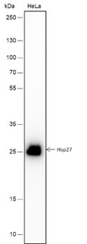
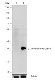
Blocking buffer: 5% NFDM/TBST, Primary Ab Dilution: 1:1000, Primary Ab incubation condition: 2 hours at room temperature, Secondary Ab: Goat Anti-Rabbit IgG H&L (HRP), Lysate: HeLa, Protein loading quantity: 20 µg, Exposure time: 60 s, Predicted MW: 23 kDa, Observed MW: 27 kDa.
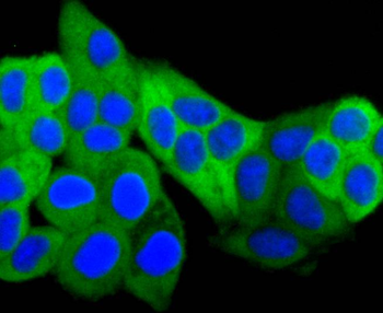
Cell line: A549, Fixative: 4% Paraformaldehyde, Permeabilization: 0.1% TritonX-100, Primary Ab Dilution: 1:300, Primary Ab incubation condition: 4°C overnight, Secondary Ab: Goat Anti-Rabbit IgG, Nuclear counter stain: DAPI (Blue), Comment: Color green is the positive signal for orb1499379.
Cell line: C2C12, Fixative: 4% Paraformaldehyde, Permeabilization: 0.1% TritonX-100, Primary Ab Dilution: 1:50, Primary Ab incubation condition: 4°C overnight, Secondary Ab: Goat Anti-Rabbit IgG, Nuclear counter stain: DAPI (Blue), Comment: Color green is the positive signal for orb1499379.
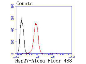
Cell line: HeLa, Fixative: 4% Paraformaldehyde, Permeabilization: 90% Methanol, Primary Ab Dilution: 1:50, Secondary Ab: Goat anti Rabbit IgG, Unlabelled control: The cell without incubation with primary antibody and secondary antibody (Black line). Isotype control: Rabbit monoclonal IgG (Blue line). Comment: Line red is the positive signal for orb1499379.
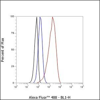
Flow cytometric analysis of Hsp27 was done on Hela cells. The cells were fixed, permeabilized and stained with the primary antibody (orb1499379, 1/50) (red). After incubation of the primary antibody at room temperature for an hour, the cells were stained with a Alexa Fluor 488-conjugated Goat anti-Rabbit IgG Secondary antibody at 1/1000 dilution for 30 minutes. Unlabelled sample was used as a control (cells without incubation with primary antibody, black).
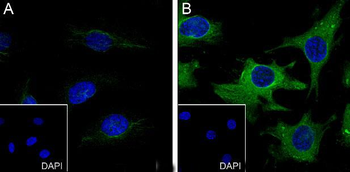
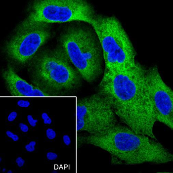
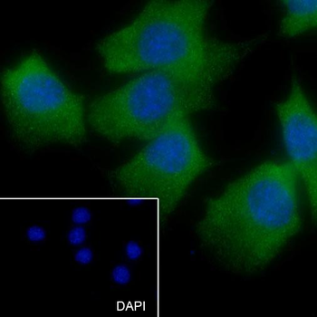
ICC staining of Hsp27 in Hela cells (green). Formalin fixed cells were permeabilized with 0.1% Triton X-100 in TBS for 10 minutes at room temperature and blocked with 1% Blocker BSA for 15 minutes at room temperature. Cells were probed with the primary antibody (orb1499379, 1/50) for 1 hour at room temperature, washed with PBS. Alexa Fluor®488 Goat anti-Rabbit IgG was used as the secondary antibody at 1/1000 dilution. The nuclear counter stain is DAPI (blue).
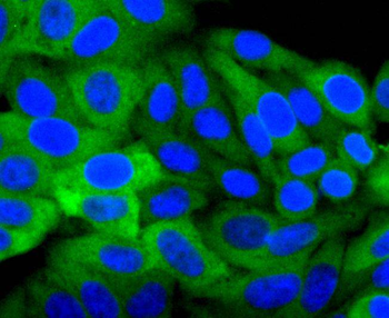
ICC staining of Hsp27 in HepG2 cells (green). Formalin fixed cells were permeabilized with 0.1% Triton X-100 in TBS for 10 minutes at room temperature and blocked with 1% Blocker BSA for 15 minutes at room temperature. Cells were probed with the primary antibody (orb1499379, 1/50) for 1 hour at room temperature, washed with PBS. Alexa Fluor®488 Goat anti-Rabbit IgG was used as the secondary antibody at 1/1000 dilution. The nuclear counter stain is DAPI (blue).
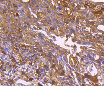
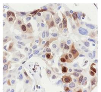
Immunohistochemical analysis of paraffin-embedded human breast carcinoma tissue using anti-Hsp27 antibody. The section was pre-treated using heat mediated antigen retrieval with Tris-EDTA buffer (pH 8.0-8.4) for 20 minutes. The tissues were blocked in 5% BSA for 30 minutes at room temperature, washed with ddH2O and PBS, and then probed with the primary antibody (orb1499379, 1/50) for 30 minutes at room temperature. The detection was performed using an HRP conjugated compact polymer system. DAB was used as the chromogen. Tissues were counterstained with hematoxylin and mounted with DPX.
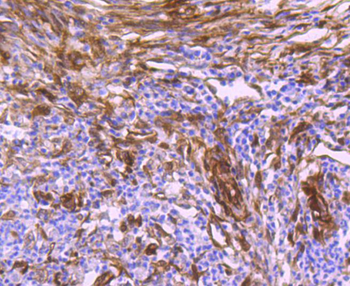
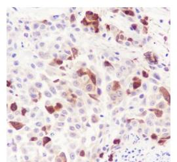
Immunohistochemical analysis of paraffin-embedded human colon carcinoma tissue using anti-Hsp27 antibody. The section was pre-treated using heat mediated antigen retrieval with Tris-EDTA buffer (pH 8.0-8.4) for 20 minutes. The tissues were blocked in 5% BSA for 30 minutes at room temperature, washed with ddH2O and PBS, and then probed with the primary antibody (orb1499379, 1/50) for 30 minutes at room temperature. The detection was performed using an HRP conjugated compact polymer system. DAB was used as the chromogen. Tissues were counterstained with hematoxylin and mounted with DPX.

Immunohistochemical analysis of paraffin-embedded human colon carcinoma tissue using anti-Hsp27 antibody. The section was pre-treated using heat mediated antigen retrieval with Tris-EDTA buffer (pH 8.0-8.4) for 20 minutes. The tissues were blocked in 5% BSA for 30 minutes at room temperature, washed with ddH2O and PBS, and then probed with the primary antibody (orb1499379, 1/50) for 30 minutes at room temperature. The detection was performed using an HRP conjugated compact polymer system. DAB was used as the chromogen. Tissues were counterstained with hematoxylin and mounted with DPX.
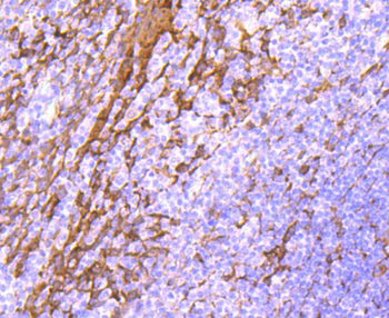
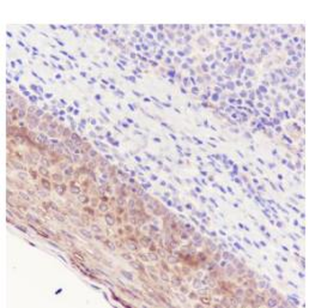
Immunohistochemical analysis of paraffin-embedded human tonsil tissue using anti-Hsp27 antibody. The section was pre-treated using heat mediated antigen retrieval with Tris-EDTA buffer (pH 8.0-8.4) for 20 minutes. The tissues were blocked in 5% BSA for 30 minutes at room temperature, washed with ddH2O and PBS, and then probed with the primary antibody (orb1499379, 1/50) for 30 minutes at room temperature. The detection was performed using an HRP conjugated compact polymer system. DAB was used as the chromogen. Tissues were counterstained with hematoxylin and mounted with DPX.
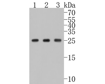
Western blot analysis of Hsp27 on different lysates. Proteins were transferred to a PVDF membrane and blocked with 5% BSA in PBS for 1 hour at room temperature. The primary antibody (orb1499379, 1/500) was used in 5% BSA at room temperature for 2 hours. Goat Anti-Rabbit IgG - HRP Secondary Antibody (HA1001) at 1:5000 dilution was used for 1 hour at room temperature. Positive control: Lane 1: Hela cell lysate, Lane 2: A549 cell lysate, Lane 3: Jurkat cell lysate.
Quick Database Links
Gene Symbol
HSPB1
UniProt
UniProt Details
− No UniProt data available
Documents Download
Datasheet
Product Information
Request a Document
Protocol Information
WB
Western Blot (IB, immunoblot)
IHC-P
Immunohistochemistry Paraffin
IHC-Fr
Immunohistochemistry Frozen
FC
Flow Cytometry
IF
Immunofluorescence
ICC
Immunocytochemistry
Hsp27 Recombinant Rabbit Monoclonal Antibody (orb1499379)

- 0.0
Based on 0 reviews
Participating in our Biorbyt product reviews program enables you to support fellow scientists by sharing your firsthand experience with our products.
Login to Submit a ReviewAvailable Sizes
Select a size below
Free Secondary Antibody (20 ul)0/0
Please add an antibody product to your cart first.