You have no items in your shopping cart.
Description
Research Area
Cancer Biology
Images & Validation
−Item 1 of 7
| Tested Applications | IF, IHC-P, WB |
|---|---|
| Dilution Range | IF - 1:10-50, WB - 1:2000, IHC-P - 1:10-50 |
| Reactivity | Human |
Key Properties
−| Host | Rabbit |
|---|---|
| Clonality | Polyclonal |
| Isotype | Rabbit IgG |
| Immunogen | This p53 antibody is generated from rabbits immunized with a KLH conjugated synthetic peptide between 293-322 amino acids from human p53. Antigen Region: 293-322 aa. |
| Target | TP53 |
| Molecular Weight | 43653 Da |
| Conjugation | Unconjugated |
Storage & Handling
−| Storage | Maintain refrigerated at 2-8°C for up to 2 weeks. For long term storage store at -20°C in small aliquots to prevent freeze-thaw cycles |
|---|---|
| Form/Appearance | Purified polyclonal antibody supplied in PBS with 0.09% (W/V) sodium azide. This antibody is purified through a protein A column, followed by peptide affinity purification. |
| Expiration Date | 12 months from date of receipt. |
| Disclaimer | For research use only |
Alternative Names
−Cellular tumor antigen p53, Antigen NY-CO-13, Phosphoprotein p53, Tumor suppressor p53, TP53, P53
Similar Products
−- Item 1 of 3
p53 (Phospho-S315) Rabbit Polyclonal Antibody [orb214692]
IF, IHC, IP, WB
Human, Monkey, Rat
Rabbit
Polyclonal
Unconjugated
30 μl, 100 μl, 200 μl, 50 μl - Item 1 of 2
Phospho-p53(S315) Antibody [orb1931242]
DOT, IHC-P, WB
Monkey
Human
Rabbit
Polyclonal
Unconjugated
50 μl, 100 μl - Item 1 of 2
Phospho-P53 (Ser315) Rabbit Polyclonal Antibody [orb6595]
FC, WB
Mouse
Human, Mouse
Rabbit
Polyclonal
Unconjugated
50 μl, 100 μl, 200 μl 
Quality Guarantee
Explore bioreagents carefree to elevate your research. All our products are rigorously tested for performance. If a product does not perform as described on its datasheet, our scientific support team will provide expert troubleshooting, a prompt replacement, or a refund. For full details, please see our Terms & Conditions and Buying Guide. Contact us at [email protected].
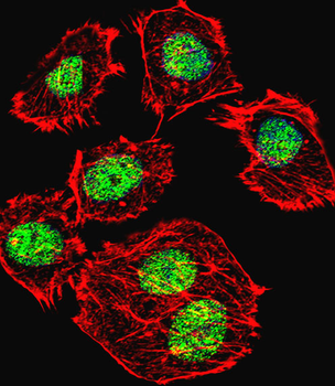
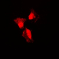
Fluorescent confocal image of U251 cell stained with p53 Antibody (S315).U251 cells were fixed with 4% PFA (20 min), permeabilized with Triton X-100 (0.1%, 10 min), then incubated with p53 primary antibody (1:25, 1 h at 37°C). For secondary antibody, Alexa Fluor 488 conjugated donkey anti-rabbit antibody (green) was used (1:400, 50 min at 37°C).Cytoplasmic actin was counterstained with Alexa Fluor 555 (red) conjugated Phalloidin (7units/ml, 1 h at 37°C). Nuclei were counterstained with DAPI (blue) (10 µg/ml, 10 min). p53 immunoreactivity is localized to Nucleus significantly.
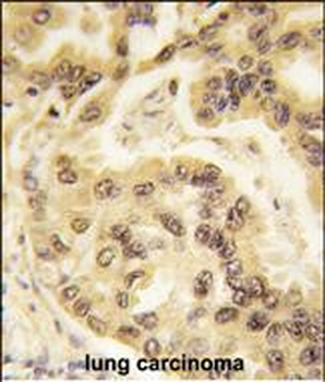
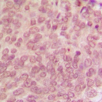
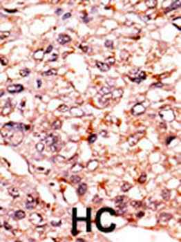
Formalin-fixed and paraffin-embedded human lung carcinoma tissue reacted with p53 Antibody (S315), which was peroxidase-conjugated to the secondary antibody, followed by DAB staining. This data demonstrates the use of this antibody for immunohistochemistry; clinical relevance has not been evaluated.
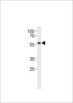
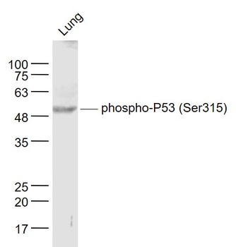
p53 Antibody (S315) western blot analysis in Daudi cell line lysates (35 ug/lane). This demonstrates the p53 antibody detected the p53 protein (arrow).
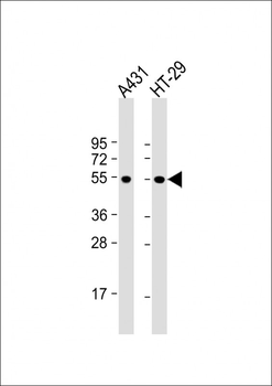
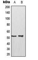
All lanes: Anti-p53 Antibody (S315) at 1:2000 dilution. Lane 1: A431 whole cell lysate. Lane 2: HT-29 whole cell lysate. Lysates/proteins at 20 µg per lane. Secondary Goat Anti-Rabbit IgG, (H+L), Peroxidase conjugated at 1/10000 dilution. Predicted band size: 44 kDa. Blocking/Dilution buffer: 5% NFDM/TBST.
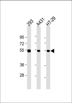
All lanes: Anti-p53 Antibody (S315) at 1:2000 dilution. Lane 1: 293 whole cell lysate. Lane 2: A431 whole cell lysate. Lane 3: HT-29 whole cell lysate. Lysates/proteins at 20 µg per lane. Secondary Goat Anti-Rabbit IgG, (H+L), Peroxidase conjugated at 1/10000 dilution. Predicted band size: 44 kDa. Blocking/Dilution buffer: 5% NFDM/TBST.
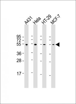
All lanes: Anti-p53 Antibody (S315) at 1:2000 dilution. Lane 1: A431 whole cell lysate. Lane 2: Hela whole cell lysate. Lane 3: HT-29 whole cell lysate. Lane 4: MCF-7 whole cell lysate. Lysates/proteins at 20 µg per lane. Secondary Goat Anti-Rabbit IgG, (H+L), Peroxidase conjugated at 1/10000 dilution. Predicted band size: 44 kDa. Blocking/Dilution buffer: 5% NFDM/TBST.
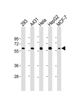
All lanes: Anti-p53 Antibody (S315) at 1:2000 dilution. Lane 1: 293 whole cell lysate. Lane 2: A431 whole cell lysate. Lane 3: Hela whole cell lysate. Lane 4: HepG2 whole cell lysate. Lane 5: MCF-7 whole cell lysate. Lysates/proteins at 20 µg per lane. Secondary Goat Anti-Rabbit IgG, (H+L), Peroxidase conjugated at 1/10000 dilution. Predicted band size: 44 kDa. Blocking/Dilution buffer: 5% NFDM/TBST.
Quick Database Links
Gene Symbol
TP53
UniProt
UniProt Details
− No UniProt data available
NCBI Reference Sequences
−Associated Accession Numbers
Curated reference sequences for the gene transcript and protein productDocuments Download
Datasheet
Product Information
Request a Document
Protocol Information
WB
Western Blot (IB, immunoblot)
IHC-P
Immunohistochemistry Paraffin
IF
Immunofluorescence
p53 Antibody (S315) (orb1930141)

- 0.0
Based on 0 reviews
Participating in our Biorbyt product reviews program enables you to support fellow scientists by sharing your firsthand experience with our products.
Login to Submit a ReviewAvailable Sizes
Select a size below
Choose Conjugation or Carrier Free Version
Free Secondary Antibody (20 ul)0/0
Please add an antibody product to your cart first.