You have no items in your shopping cart.
Description
Images & Validation
−Item 1 of 8
| Tested Applications | FC, ICC, IF, IHC, WB |
|---|---|
| Dilution Range | WB: 1:1,000-1:2,000 FC: 1:50-1:100 ICC/IF: 1:50-1:200 IHC: 1:50-1:200 |
| Reactivity | Human, Mouse, Rat |
Key Properties
−| Antibody Type | Primary Antibody |
|---|---|
| Host | Rabbit |
| Clonality | Recombinant |
| Clone No. | BLI62X28 |
| Immunogen | Recombinant protein within human SNX1 aa 50-250. |
| Molecular Weight | Calculated MW 63 kDa |
| Purity | ProA affinity purified. |
| Conjugation | Unconjugated |
Storage & Handling
−| Storage | Store at -20˚C. |
|---|---|
| Form/Appearance | Liquid |
| Buffer/Preservatives | 1*TBS (pH7.4), 1% rAlbumin, 40% Glycerol, 0.05% Sodium Azide |
| Expiration Date | 12 months from date of receipt. |
| Disclaimer | For research use only |
Alternative Names
−HsT17379 antibody/ MGC8664 antibody/ SNX 1 antibody/ SNX 1a antibody/ Snx1 antibody/ SNX1_HUMAN antibody/ SNX1A antibody/ Sorting nexin 1 antibody/ Sorting nexin 1A antibody/ Sorting nexin-1 antibody/ Vps5 antibody
Similar Products
−- Item 1 of 6
SNX1 Rabbit Polyclonal Antibody [orb1474817]
ELISA, FC, ICC, IF, IHC, WB
Human
Rabbit
Polyclonal
Unconjugated
100 μg - Item 1 of 4
SNX1 Antibody [orb20425]
ELISA, ICC, IHC, WB
Bovine, Canine, Mouse, Rat
Human
Goat
Polyclonal
Unconjugated
100 μg - Item 1 of 2
SNX1 rabbit pAb Antibody [orb766350]
ELISA, IHC, WB
Human, Mouse, Rat
Polyclonal
Unconjugated
100 μl, 50 μl - Item 1 of 3
SNX1 Rabbit Polyclonal Antibody [orb331258]
WB
Bovine, Canine, Equine, Guinea pig, Mouse, Rabbit, Rat
Human
Rabbit
Polyclonal
Unconjugated
100 μl - Item 1 of 3
SNX1 Antibody [orb1247188]
ELISA, ICC, IHC, WB
Bovine, Canine, Mouse, Rat
Human
Goat
Polyclonal
Unconjugated
0.1 mg


Quality Guarantee
Explore bioreagents carefree to elevate your research. All our products are rigorously tested for performance. If a product does not perform as described on its datasheet, our scientific support team will provide expert troubleshooting, a prompt replacement, or a refund. For full details, please see our Terms & Conditions and Buying Guide. Contact us at [email protected].
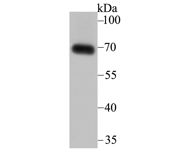
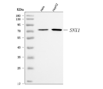
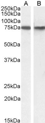
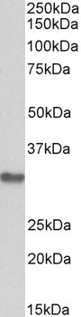
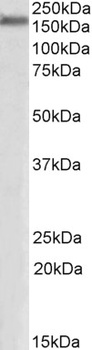
Western blot analysis of SNX1 on human skin tissue lysate using anti-SNX1 antibody at 1/2000 dilution
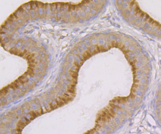
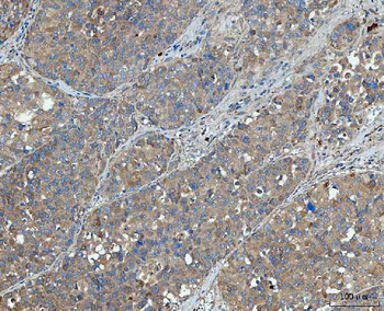
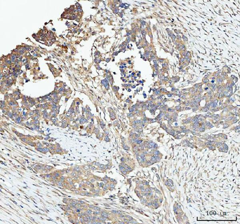
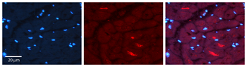
Immunohistochemical analysis of paraffin-embedded rat epididymis tissue using anti-SNX1 antibody. Counter stained with hematoxylin
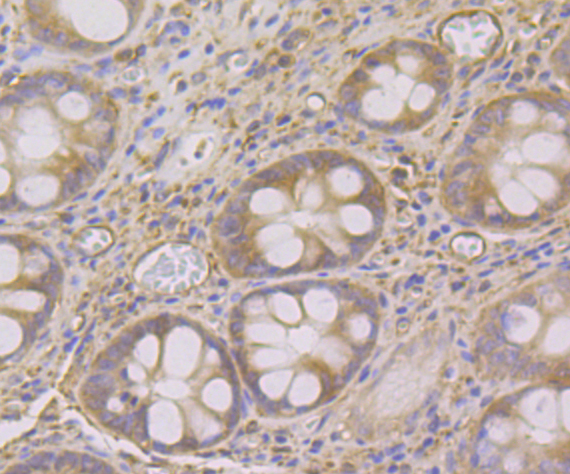
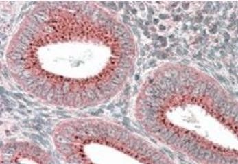
Immunohistochemical analysis of paraffin-embedded human colon tissue using anti-SNX1 antibody. Counter stained with hematoxylin
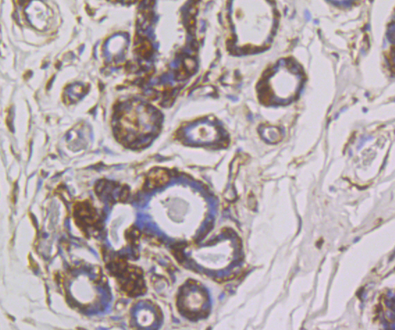
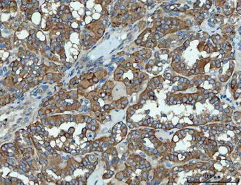
Immunohistochemical analysis of paraffin-embedded human breast cancer tissue using anti-SNX1 antibody. Counter stained with hematoxylin
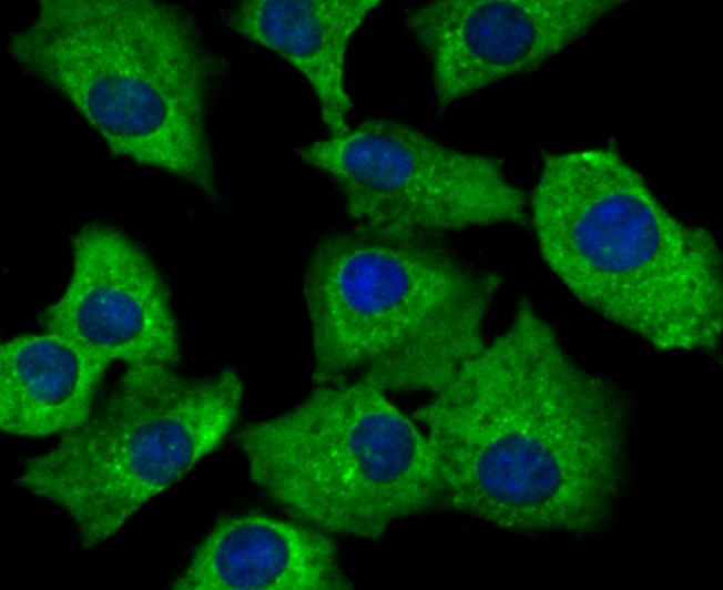
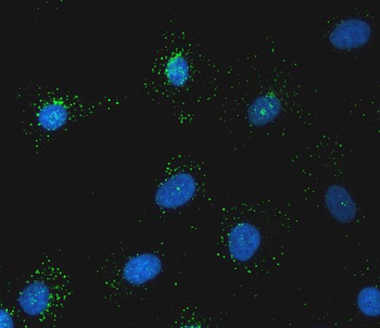
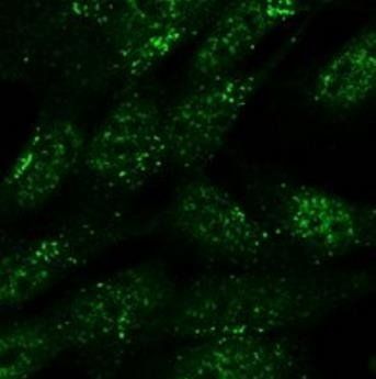
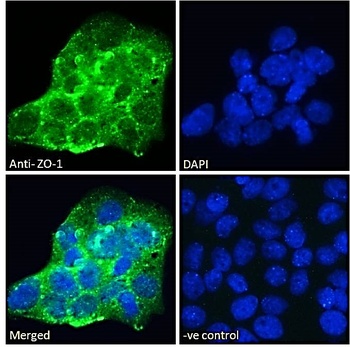
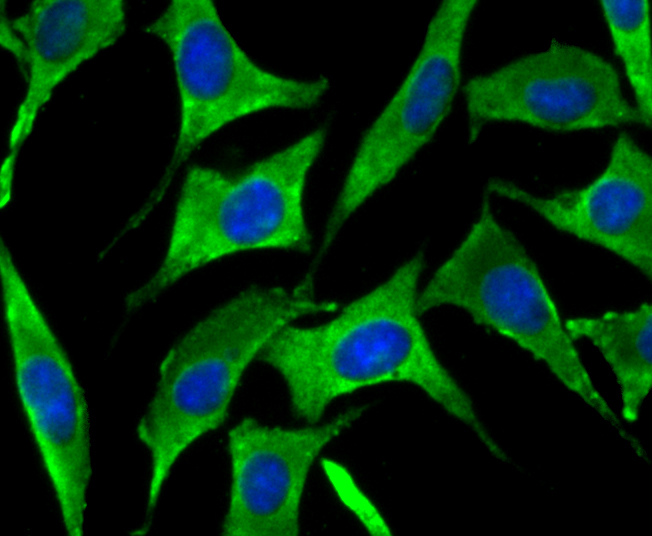
ICC staining SNX1 in A549 cells (green). The nuclear counter stain is DAPI (blue). Cells were fixed in paraformaldehyde, permeabilised with 0.25% Triton X100/PBS
ICC staining SNX1 in SH-SY-5Y cells (green). The nuclear counter stain is DAPI (blue). Cells were fixed in paraformaldehyde, permeabilised with 0.25% Triton X100/PBS
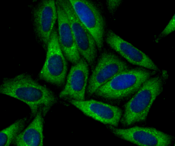
ICC staining SNX1 in SiHa cells (green). The nuclear counter stain is DAPI (blue). Cells were fixed in paraformaldehyde, permeabilised with 0.25% Triton X100/PBS
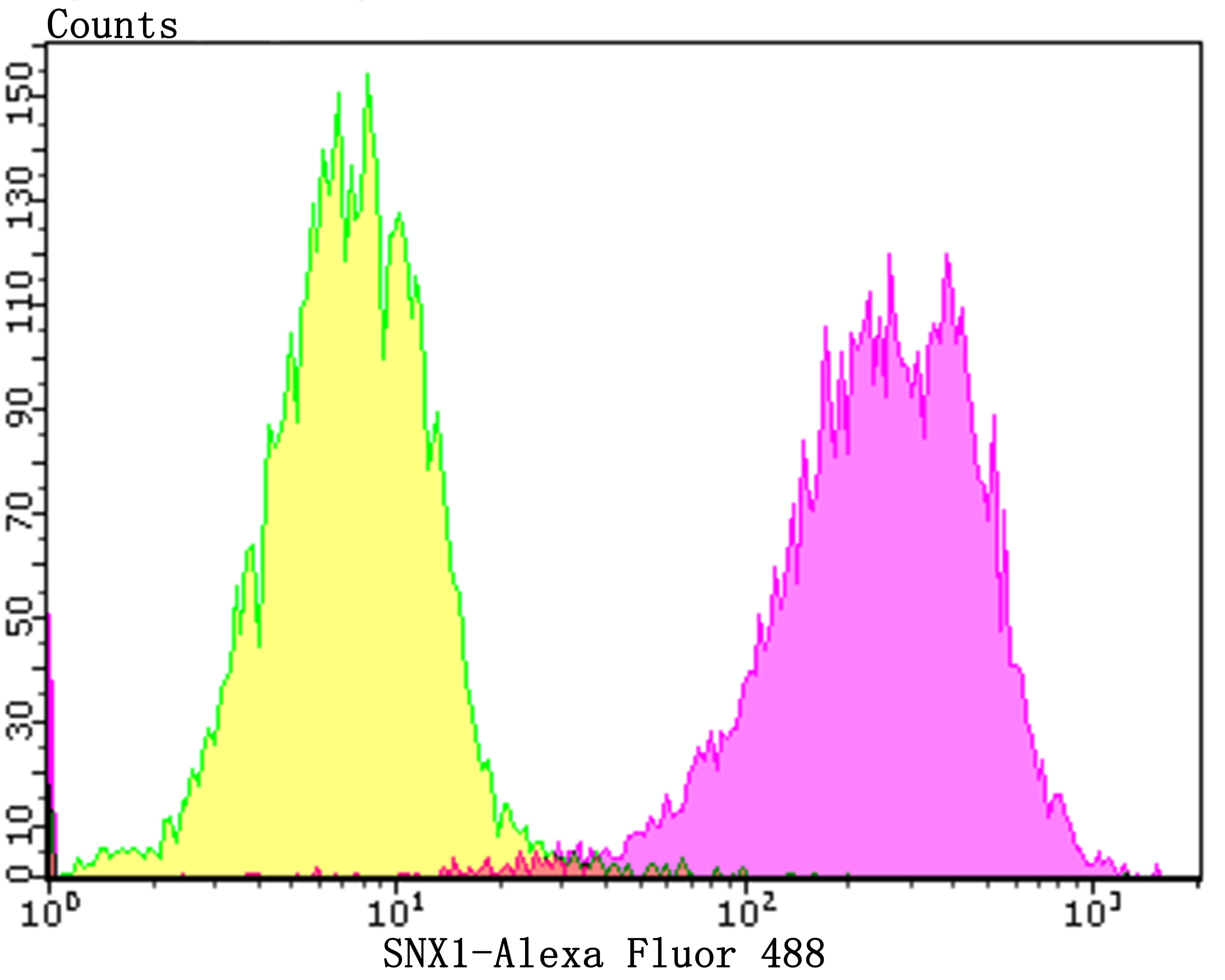
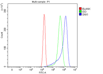
Flow cytometric analysis of SiHa cells with SNX1 antibody at 1/100 dilution (purple) compared with an unlabelled control (cells without incubation with primary antibody; yellow). Alexa Fluor 488-conjugated goat anti-rabbit IgG was used as the secondary antibody
Quick Database Links
UniProt
UniProt Details
− No UniProt data available
Documents Download
Datasheet
Product Information
Request a Document
Protocol Information
WB
Western Blot (IB, immunoblot)
IHC
Immunohistochemistry
FC
Flow Cytometry
IF
Immunofluorescence
ICC
Immunocytochemistry
SNX1 Antibody (orb622711)

- 0.0
Based on 0 reviews
Participating in our Biorbyt product reviews program enables you to support fellow scientists by sharing your firsthand experience with our products.
Login to Submit a ReviewAvailable Sizes
Select a size below
Choose Conjugation or Carrier Free Version
Free Secondary Antibody (20 ul)0/0
Please add an antibody product to your cart first.