
You have no items in your shopping cart.
Immunoassays & Diagnostics
Streptavidin-Biotin System
A core signal amplification platform enabling unparalleled sensitivity and precise detection in the fields of life science research and in vitro diagnostics.
1. Biological Overview
In the fields of life science research and in vitro diagnostics, the streptavidin-biotin system stands as an indispensable core technology platform. Leveraging the near-irreversible non-covalent interaction between these two molecules (with a dissociation constant, Kd ≈ 10-15 M), this system enables efficient signal amplification and precise detection. It is widely applied in immunology, molecular biology, cell imaging, targeted delivery, and numerous other areas.
Its exceptional sensitivity and stability are primarily attributed to the unique properties of its core components—biotin and streptavidin—and their remarkably specific interaction.
2. Biotin: The Universal Connector
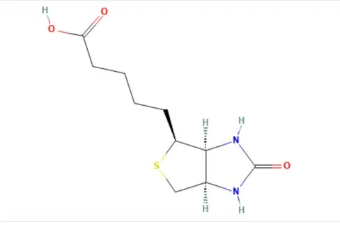
Biotin, also known as Vitamin H, is a water-soluble small molecule vitamin. Its molecular structure features a sulfur-containing thiophene ring, to which a ureido ring and a valeric acid side chain are attached. This compact and stable architecture is fundamental to its function.
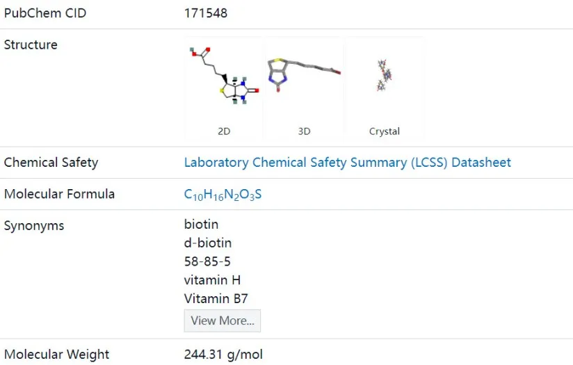
Chemical structure of Biotin (Source: PubChem)
The carboxyl group (-COOH) of the valeric acid side chain can be readily activated, allowing biotin to be covalently conjugated to various molecules such as proteins (e.g., antibodies), nucleic acids, drugs, or fluorescent dyes, with minimal impact on its ability to bind streptavidin. This makes biotin an ideal "universal tag" or "linking bridge."
Schematic of biotin as a universal tag conjugated to a target molecule
3. Streptavidin: The High-Performance Capture Protein
Streptavidin (SA), derived from Streptomyces avidinii, is a homotetrameric protein. Each of its four subunits can independently bind one biotin molecule with exceptionally high affinity. Compared to another biotin-binding protein, avidin (from egg white), streptavidin offers significant advantages:
- Streptavidin: Features a near-neutral isoelectric point and is non-glycosylated, resulting in extremely low non-specific binding under physiological conditions.
- Avidin: Is a basic glycoprotein that tends to bind non-specifically to negatively charged structures like cell membranes, leading to higher background signals.
Recombinantly expressed streptavidin can be easily and stably fused or conjugated with a wide range of reporter molecules (e.g., horseradish peroxidase HRP, fluorescein FITC, phycoerythrin PE, alkaline phosphatase AP), creating ready-to-use detection tools.
4. The Extraordinary Binding Strength
The remarkable affinity of the streptavidin-biotin system arises from the precise steric complementarity and multiple synergistic interactions between biotin and the streptavidin binding pocket.
Each subunit of streptavidin possesses a binding site exquisitely matched to biotin, with a spatial structure highly complementary to it. Upon entry into this site, the molecular surfaces of biotin and streptavidin fit tightly together, establishing multiple interaction points that enable specific recognition and binding.
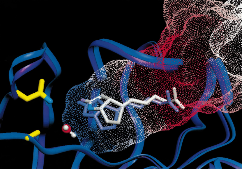
The association is driven by a combination of non-covalent interactions, including hydrogen bonds, van der Waals forces, and hydrophobic effects. These interactions work cooperatively to form an exceptionally stable complex. For instance, nitrogen atoms in biotin's ureido and imidazolidone rings form hydrogen bonds with amino acid residues in the streptavidin subunit, while biotin's valeric acid side chain engages in hydrophobic interactions with hydrophobic residues lining the binding pocket.
Schematic diagram of the biotin dissociation pathway derived from PMF simulations, superimposed on the D128A crystal structure
5. Multiple Advantages of the System
Based on these biochemical characteristics, the streptavidin-biotin system offers numerous robust advantages for modern immunoassays:
- Signal Amplification: A single biotinylated target molecule can bind multiple labeled streptavidin molecules, significantly enhancing the detection signal for low-abundance targets.
- High Flexibility: Biotin can be conjugated to diverse molecules, and streptavidin is available in various labeled formats, making it adaptable to a wide array of techniques including ELISA, Western Blot (WB), Immunohistochemistry (IHC), Flow Cytometry (FACS), and Fluorescence in Situ Hybridization (FISH).
- Low Background & High Signal-to-Noise Ratio: The low non-specific binding characteristic of streptavidin ensures high specificity and exceptionally clean background in detection assays.
- Robust Stability: The formed complex withstands a broad range of pH, temperature, organic solvents, and protein denaturants, ensuring excellent experimental reproducibility and tolerance.
6. A Complete One-Stop Detection Solution
Biorbyt provides highly validated streptavidin conjugate reagents engineered for precision and rapid signal processing across major life science research methodologies.
| Product ID | Description & Application |
|---|---|
| orb86224 |
Streptavidin, HRP conjugated
Chemiluminescent / Colorimetric Detection
The core tool for high-sensitivity detection. Precisely guides the HRP enzyme to any biotinylated target. Widely used in ELISA, WB, and IHC systems requiring maximum sensitivity utilizing substrates like TMB, DAB, and ECL. |
| orb1878761 |
Streptavidin-FITC
Green Fluorescence Labeling
A versatile visualization tool providing a precise green fluorescent signal to biotin-labeled sites. Applied in immunofluorescence staining (IF/IHC), ELISA, and Flow Cytometry. (Ex/Em ~494/519 nm). |
| orb2321154 |
Streptavidin PE
Flow Cytometry Analysis
A gold-standard tool for ultra-bright flow cytometry. PE efficiently directs to the target via the biotin-streptavidin bridge. An indispensable reagent for multi-color flow cytometry, producing intense orange-red fluorescence (Ex/Em ~565/578 nm). |
| orb1671193 |
Streptavidin (Alkaline Phosphatase)
Stable Chromogenic Detection
Anchors the AP enzyme to the target, catalyzing substrates to produce stable colorimetric signals. Particularly advantageous in systems containing HRP inhibitors or requiring long development times (e.g., BCIP/NBT, PNPP). |
7. Comprehensive Complementary Products
Alongside our core Streptavidin-Biotin solutions, Biorbyt offers an extensive range of supporting detection reagents to streamline your workflow and expand your analytical capabilities.
Enzyme-Labeled Antibodies
Designed for ultra-high sensitivity signal amplification. Includes HRP conjugated antibodies ideal for ELISA, Western Blot, and IHC, as well as AP conjugated options for robust membrane-based assays and excelling with specific chromogenic substrates.
Fluorophore-Labeled Antibodies
Essential for multi-color imaging and flow cytometry analysis. Features classic fluorophores including FITC (green), PE (bright orange-red), and APC (far-red), plus next-generation dyes like Alexa Fluor® and Cy® series for enhanced photostability.
Biotinylated Antibodies
A vast, highly-validated selection covering thousands of distinct biological targets. This portfolio spans heavily researched primary antibodies as well as versatile, multi-species secondary antibodies (e.g., Biotinylated Goat Anti-Mouse IgG).
8. References
- https://www.sciencedirect.com/science/article/abs/pii/S1093326317300943
- Stayton PS, Freitag S, Klumb LA, Chilkoti A, Chu V, Penzotti JE, To R, Hyre D, Le Trong I, Lybrand TP, Stenkamp RE. Streptavidin-biotin binding energetics. Biomol Eng. 1999 Dec 31;16(1-4):39-44. doi: 10.1016/s1050-3862(99)00042-x. PMID: 10796983.