You have no items in your shopping cart.
Description
Research Area
Immunology & Inflammation
Images & Validation
−Item 1 of 3
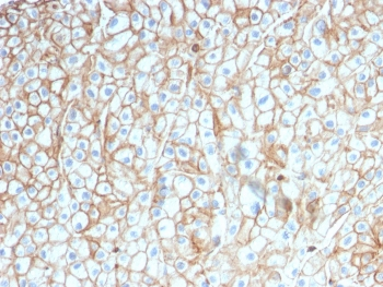
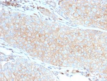
| Tested Applications | FC, ICC, IHC-Fr, IP, WB |
|---|---|
| Reactivity | Human, Primate |
| Application Notes |
Key Properties
−| Antibody Type | Primary Antibody |
|---|---|
| Clonality | Monoclonal |
| Isotype | Mouse IgG1 kappa |
| Clone No. | CD28.2 |
| Immunogen | DC28.1.3.3 murine T cell hybridoma transfected with human CD28 cDNA |
| Target | CD28 |
| Purification | Purified by protein-A affinity chromatography. |
| Conjugation | Unconjugated |
Storage & Handling
−| Storage | Maintain refrigerated at 2-8°C for up to 2 weeks. For long term storage store at -20°C in small aliquots to prevent freeze-thaw cycles. |
|---|---|
| Buffer/Preservatives | Phosphate buffered saline (PBS), pH 7.4, 15 mM sodium azide |
| Concentration | 1 mg/ml |
| Expiration Date | 12 months from date of receipt. |
| Disclaimer | For research use only |
Alternative Names
−TP44
Similar Products
−- Item 1 of 11
CD28 Rabbit Polyclonal Antibody [orb378206]
ICC, IF, IHC-P, WB
Human, Mouse, Rat
Rabbit
Polyclonal
Unconjugated
100 μg - Item 1 of 3
CD86 Antibody [orb388891]
IF, IHC, WB
Human, Mouse, Rat
Mouse
Monoclonal
Unconjugated
20 μg, 100 μg, 100 μg (without BSA and Azide) - Item 1 of 10
PD-L1 Antibody / B7-H1 / CD274 [orb2641418]
ELISA, FACS, IF, IHC-P, WB
Human, Mouse
Mouse
Monoclonal
Unconjugated
100 μg - Item 1 of 10
PD-L1 Antibody / B7-H1 / CD274 [orb606675]
ELISA, FACS, IF, IHC-P, WB
Human, Mouse
Mouse
Monoclonal
Unconjugated
100 μg, 20 μg - Item 1 of 7


Quality Guarantee
Explore bioreagents carefree to elevate your research. All our products are rigorously tested for performance. If a product does not perform as described on its datasheet, our scientific support team will provide expert troubleshooting, a prompt replacement, or a refund. For full details, please see our Terms & Conditions and Buying Guide. Contact us at [email protected].
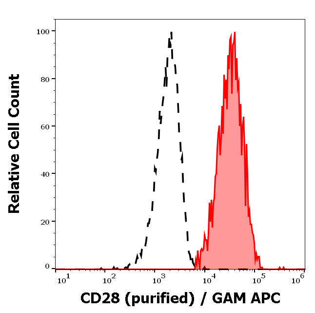
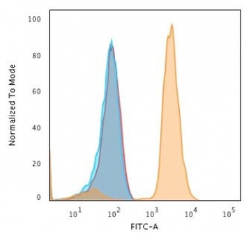
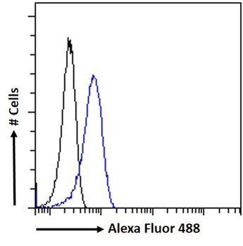
Separation of human CD28 positive lymphocytes (red-filled) from neutrophil granulocytes (black-dashed) in flow cytometry analysis (surface staining) of human peripheral whole blood stained using anti-human CD28 (CD28.2) purified antibody (concentration in sample 1.6 µg/ml) GAM APC.
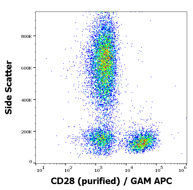
Flow cytometry surface staining pattern of human peripheral blood stained using anti-human CD28 (CD28.2) purified antibody (concentration in sample 1.6 µg/ml) GAM APC.
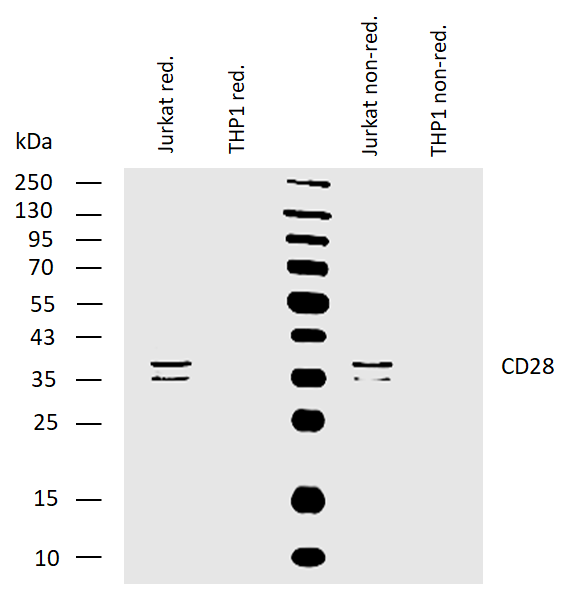
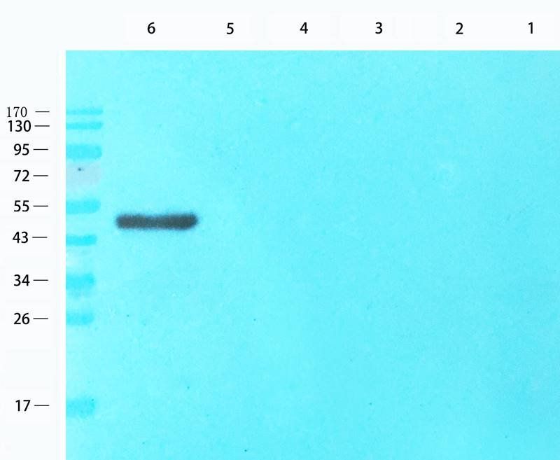
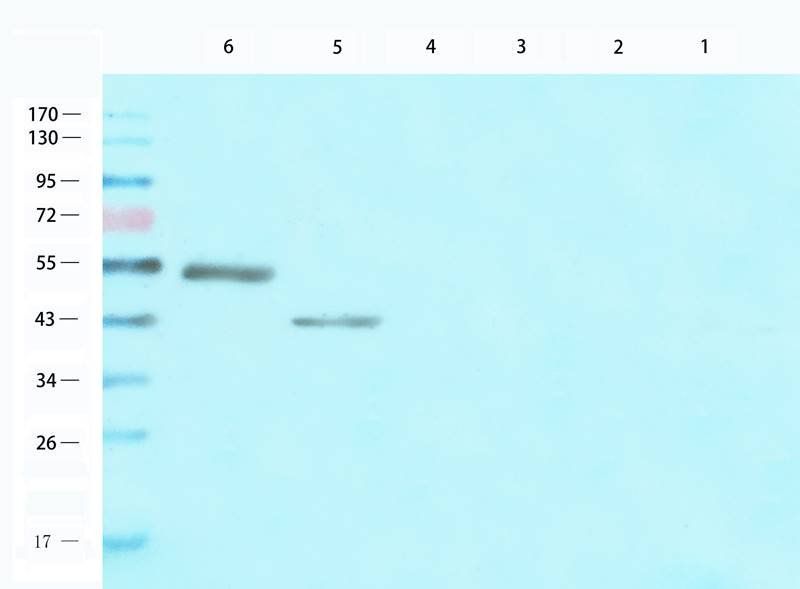
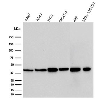
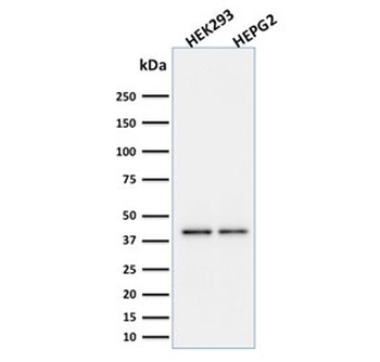
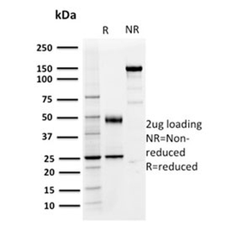
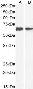
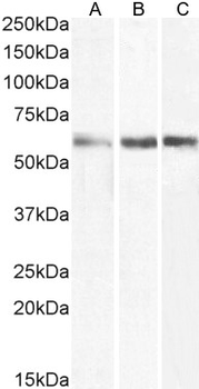
Western blotting analysis of human CD28 using mouse monoclonal antibody CD28.2 on lysates of Jurkat cells and THP1 cells (negative control) under reducing and non-reducing conditions. Nitrocellulose membrane was probed with 2 µg/ml of mouse monoclonal antibody followed by IRDye800-conjugated anti-mouse secondary antibody. CD28 was detected around 35-40 kDa.
Documents Download
Datasheet
Product Information
Request a Document
Protocol Information
WB
Western Blot (IB, immunoblot)
IHC-Fr
Immunohistochemistry Frozen
FC
Flow Cytometry
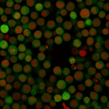
ICC
Immunocytochemistry
IP
Immunoprecipitation
CD28 Antibody (orb43723)

- 0.0
Based on 0 reviews
Participating in our Biorbyt product reviews program enables you to support fellow scientists by sharing your firsthand experience with our products.
Login to Submit a ReviewAvailable Sizes
Select a size below
Free Secondary Antibody (20 ul)0/0
Please add an antibody product to your cart first.