You have no items in your shopping cart.
Description
Research Area
Immunology & Inflammation
Images & Validation
−Item 1 of 3
| Tested Applications | FC, ICC |
|---|---|
| Reactivity | Human |
| Application Notes |
Key Properties
−| Antibody Type | Primary Antibody |
|---|---|
| Clonality | Monoclonal |
| Isotype | Mouse IgG2b kappa |
| Clone No. | TB3 |
| Target | CD3 |
| Purification | Purified by protein-A affinity chromatography. |
| Conjugation | Unconjugated |
Storage & Handling
−| Storage | Maintain refrigerated at 2-8°C for up to 2 weeks. For long term storage store at -20°C in small aliquots to prevent freeze-thaw cycles. |
|---|---|
| Buffer/Preservatives | Phosphate buffered saline (PBS), pH 7.4, 15 mM sodium azide |
| Concentration | 1 mg/ml |
| Expiration Date | 12 months from date of receipt. |
| Disclaimer | For research use only |
Alternative Names
−CD3E, T3E, TCRE
Similar Products
−- Item 1 of 10
CD3 Rabbit Polyclonal Antibody [orb348965]
IHC-P, WB
Human, Mouse, Rat
Rabbit
Polyclonal
Unconjugated
100 μg - Item 1 of 8
CD3 epsilon/CD3E Rabbit Polyclonal Antibody [orb196263]
ICC, IF, IHC, IHC-Fr, WB
Gallus, Human, Mouse, Rat
Rabbit
Polyclonal
Unconjugated
100 μg - Item 1 of 8
CD3 Intracellular domain Rabbit Polyclonal Antibody [orb1294242]
IF, IHC, WB
Human, Mouse, Rat
Rabbit
Polyclonal
Unconjugated
100 μl, 25 μl - Item 1 of 5
- Item 1 of 6
CD3E Rabbit Polyclonal Antibody [orb378203]
IHC-P
Human, Mouse, Rat
Rabbit
Polyclonal
Unconjugated
100 μg


![Anti-CD3 epsilon [YTH 12.5]](/images/pub/media/catalog/product/NewWebsite/35/orb411552_1.png)
![Anti-CD3 epsilon [YTH 12.5]](/images/pub/media/catalog/product/NewWebsite/35/orb411552_2.png)
![Anti-CD3 epsilon [YTH 12.5]](/images/pub/media/catalog/product/NewWebsite/35/orb411552_3.png)
![Anti-CD3 epsilon [YTH 12.5]](/images/pub/media/catalog/product/NewWebsite/35/orb411552_4.png)
![Anti-CD3 epsilon [YTH 12.5]](/images/pub/media/catalog/product/NewWebsite/35/orb411552_5.png)


Quality Guarantee
Explore bioreagents carefree to elevate your research. All our products are rigorously tested for performance. If a product does not perform as described on its datasheet, our scientific support team will provide expert troubleshooting, a prompt replacement, or a refund. For full details, please see our Terms & Conditions and Buying Guide. Contact us at [email protected].
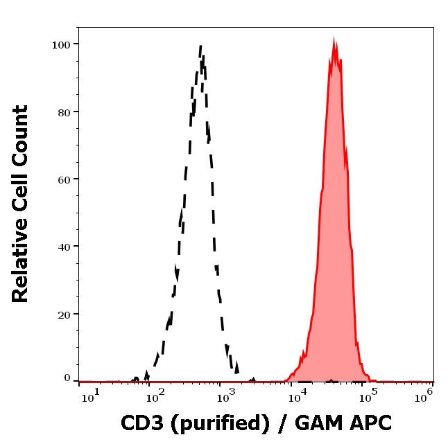
Separation of human CD3 positive lymphocytes (red-filled) from neutrophil granulocytes (black-dashed) in flow cytometry analysis (surface staining) of human peripheral whole blood stained using anti-human CD3 (TB3) purified antibody (concentration in sample 0.3 μg/ml) GAM APC.
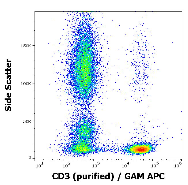
Flow cytometry surface staining pattern of human peripheral whole blood stained using anti-human CD3 (TB3) purified antibody (concentration in sample 0.3 μg/ml) GAM APC.
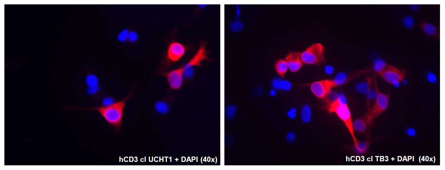
Immunocytochemistry staining of human CD3 epsilon and CD3 gamma transfected COS cells using mouse monoclonal antibodies anti-CD3, clones UCHT1 (left) and TB3 (right) indicated by Alexa Fluor 555 signal (red); DNA stained by DAPI (blue).
Documents Download
Datasheet
Product Information
Request a Document
Protocol Information
FC
Flow Cytometry
ICC
Immunocytochemistry
CD3 Antibody (orb421969)

- 0.0
Based on 0 reviews
Participating in our Biorbyt product reviews program enables you to support fellow scientists by sharing your firsthand experience with our products.
Login to Submit a ReviewAvailable Sizes
Select a size below
Free Secondary Antibody (20 ul)0/0
Please add an antibody product to your cart first.