You have no items in your shopping cart.
CD54 Antibody
SKU: orb44030
Description
Research Area
Epigenetics, Infectious Diseases
Images & Validation
−Item 1 of 5
| Tested Applications | ELISA, FC, ICC, IHC-P, WB |
|---|---|
| Reactivity | Bovine, Human, Rat |
| Application Notes |
Key Properties
−| Antibody Type | Primary Antibody |
|---|---|
| Clonality | Monoclonal |
| Isotype | Mouse IgG2a |
| Clone No. | MEM-111 |
| Immunogen | Raji human Burkitt's lymphoma cell line |
| Target | CD54 |
| Purification | Purified by protein-A affinity chromatography. |
| Conjugation | Unconjugated |
Storage & Handling
−| Storage | Maintain refrigerated at 2-8°C for up to 2 weeks. For long term storage store at -20°C in small aliquots to prevent freeze-thaw cycles. |
|---|---|
| Buffer/Preservatives | Phosphate buffered saline (PBS), pH 7.4, 15 mM sodium azide |
| Concentration | 1 mg/ml |
| Expiration Date | 12 months from date of receipt. |
| Disclaimer | For research use only |
Alternative Names
−ICAM-1, BB2, P3.58
Similar Products
−- Item 1 of 9
ICAM1 Rabbit Polyclonal Antibody [orb402187]
ELISA, FC, ICC, IF, IHC, WB
Human, Mouse, Rat
Rabbit
Polyclonal
Unconjugated
100 μg - Item 1 of 5
ICAM1 Rabbit Polyclonal Antibody [orb339615]
IHC-P, WB
Mouse, Rat
Rabbit
Polyclonal
Unconjugated
100 μg, 500 μg - Item 1 of 6
- Item 1 of 4
ICAM1 Rabbit Polyclonal Antibody [orb10334]
IF, IHC-Fr, IHC-P, WB
Rat
Human
Rabbit
Polyclonal
Unconjugated
50 μl, 100 μl, 200 μl - Item 1 of 4
Goat anti-ICAM1 (aa313-327) Antibody [orb233668]
ELISA, FC, IF, WB
Human
Goat
Polyclonal
Unconjugated
100 μg


Quality Guarantee
Explore bioreagents carefree to elevate your research. All our products are rigorously tested for performance. If a product does not perform as described on its datasheet, our scientific support team will provide expert troubleshooting, a prompt replacement, or a refund. For full details, please see our Terms & Conditions and Buying Guide. Contact us at [email protected].

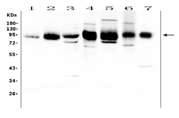
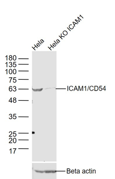
Western blotting analysis of CD54 expression in TNF-alpha activated (A) and nonactivated (B) HUVEC cells by antibody MEM-111. Lower bands represent tubulin as a loading control.

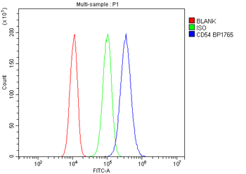
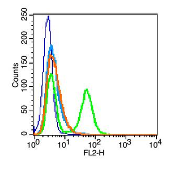
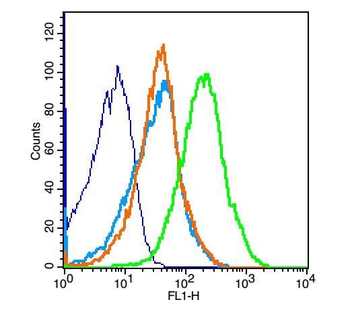
Flow cytometry multicolor surface staining pattern of human peripheral blood mononuclear cells using anti-human CD14 (MEM-15) PE antibody (20 µl reagent / 100 µl of peripheral whole blood) and anti-human CD54 (MEM-111) purified antibody (concentration in sample 0.6 µg/ml, GAM APC).
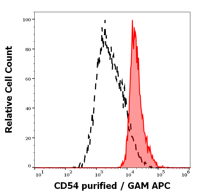
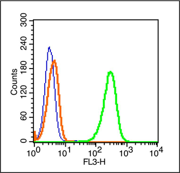
Separation of human monocytes (red-filled) from human lymphocytes (black-dashed) in flow cytometry analysis (surface staining) of human peripheral blood stained using anti-human CD54 (MEM-111) purified antibody (concentration in sample 0.6 µg/ml, GAM APC).
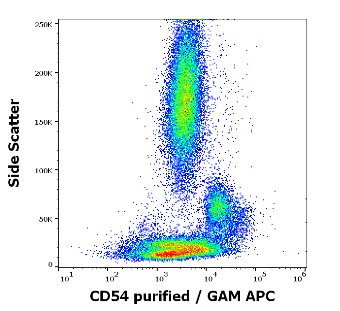
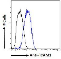
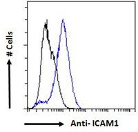
Flow cytometry surface staining pattern of human peripheral blood stained using anti-human CD54 (MEM-111) purified antibody (concentration in sample 0.6 µg/ml, GAM APC).

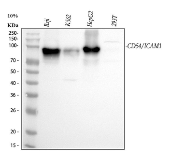
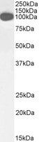
Western blotting analysis of human CD54 using mouse monoclonal antibody MEM-111 on lysates of TF-1 and Raji cells, as well as of JURKAT cells (negative control) under reducing and non-reducing conditions. Nitrocellulose membrane was probed with 2 µg/ml of mouse anti-CD54 monoclonal antibody followed by IRDye800-conjugated anti-mouse secondary antibody. A specific band was detected for CD54 at approximately 88 kDa.
Documents Download
Datasheet
Product Information
Request a Document
Protocol Information
WB
Western Blot (IB, immunoblot)
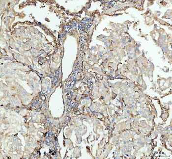
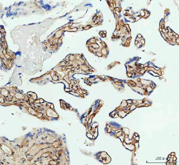
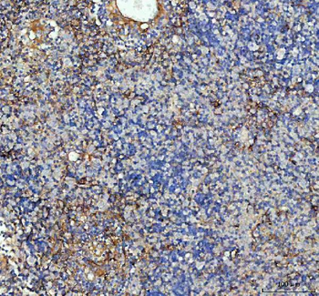
IHC-P
Immunohistochemistry Paraffin
FC
Flow Cytometry
ICC
Immunocytochemistry
ELISA
Enzyme-linked Immunosorbent Assay (EIA)
CD54 Antibody (orb44030)

- 0.0
Based on 0 reviews
Participating in our Biorbyt product reviews program enables you to support fellow scientists by sharing your firsthand experience with our products.
Login to Submit a ReviewAvailable Sizes
Select a size below