You have no items in your shopping cart.
Description
Research Area
Cancer Biology, Cell Biology, Epigenetics & Chromatin, Signal Transduction
Images & Validation
−Item 1 of 3
| Tested Applications | ICC, IF, IHC |
|---|---|
| Dilution Range | WB (1:2000), ICC/IF (1:200) |
| Reactivity | Human |
| Application Notes |
Key Properties
−| Host | Rabbit |
|---|---|
| Clonality | Polyclonal |
| Immunogen | Native human Cdc37, full length |
| Target | CDC37 |
| Molecular Weight | 44.5kDa |
| Purification | Protein A Purified |
| Conjugation | HRP |
Storage & Handling
−| Storage | Conjugated antibodies should be stored according to the product label |
|---|---|
| Buffer/Preservatives | 73.64mM Carbonate, 54.55mM Ethanolamine, 45.45mM Cyanoborohydride, 18.18mM Sodium Hydroxide, 0.23mM Citrate |
| Concentration | 1.68 mg/ml |
| Expiration Date | 12 months from date of receipt. |
| Disclaimer | For research use only |
Alternative Names
−CDC37, Hsp90 co-chaperone Cdc37, Cell division control protein 37, Hsp90 chaperone protein kinase-targeting subunit, CDC37_HUMAN, CDC37A, p50, p50Cdc37, S. cerevisiae hypothetical protein CDC37
Similar Products
−
CDC37 Rabbit Polyclonal Antibody (HRP) [orb480815]
IHC-Fr, IHC-P, WB
Bovine, Canine, Porcine, Rat
Human, Mouse
Rabbit
Polyclonal
HRP
100 μlPhospho-CDC37 (Ser13) Rabbit Polyclonal Antibody (HRP) [orb503715]
IHC-Fr, IHC-P
Bovine, Canine, Equine, Gallus, Human, Mouse, Rabbit, Rat, Sheep
Rabbit
Polyclonal
HRP
100 μl
Quality Guarantee
Explore bioreagents carefree to elevate your research. All our products are rigorously tested for performance. If a product does not perform as described on its datasheet, our scientific support team will provide expert troubleshooting, a prompt replacement, or a refund. For full details, please see our Terms & Conditions and Buying Guide. Contact us at [email protected].
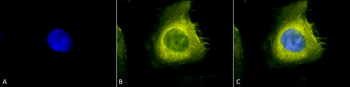
Immunocytochemistry/Immunofluorescence analysis using Rabbit Anti-CDC37 Polyclonal Antibody. Tissue: Heat Shocked Cervical cancer cell line (HeLa). Species: Human. Fixation: 2% Formaldehyde for 20 min at RT. Primary Antibody: Rabbit Anti-CDC37 Polyclonal Antibody at 1:200 for 12 hours at 4°C. Secondary Antibody: R-PE Goat Anti-Rabbit (yellow) at 1:200 for 2 hours at RT. Counterstain: DAPI (blue) nuclear stain at 1:40000 for 2 hours at RT. Localization: Cytoplasm. Magnification: 100x. (A) DAPI (blue) nuclear stain. (B) Anti-CDC37 Antibody. (C) Composite. Heat Shocked at 42°C for 30 min.
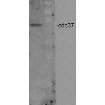
Western blot analysis of Human Cervical cancer cell line (HeLa) lysate showing detection of CDC37 protein using Rabbit Anti-CDC37 Polyclonal Antibody. Primary Antibody: Rabbit Anti-CDC37 Polyclonal Antibody at 1:2000.
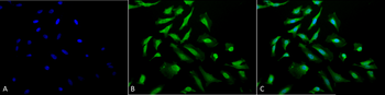
Immunocytochemistry/Immunofluorescence analysis using Rabbit Anti-CDC37 Polyclonal Antibody. Tissue: Heat Shocked Cervical cancer cell line (HeLa). Species: Human. Fixation: 2% Formaldehyde for 20 min at RT. Primary Antibody: Rabbit Anti-CDC37 Polyclonal Antibody at 1:200 for 12 hours at 4°C. Secondary Antibody: FITC Goat Anti-Rabbit (green) at 1:200 for 2 hours at RT. Counterstain: DAPI (blue) nuclear stain at 1:40000 for 2 hours at RT. Localization: Cytoplasm. Magnification: 20x. (A) DAPI (blue) nuclear stain. (B) Anti-CDC37 Antibody. (C) Composite. Heat Shocked at 42°C for 30 min.
Quick Database Links
UniProt Details
− No UniProt data available
NCBI Gene Details
− No NCBI Gene data available
NCBI Reference Sequences
−Associated Accession Numbers
Curated reference sequences for the gene transcript and protein product| Protein | NP_008996.1 |
|---|
Documents Download
Datasheet
Product Information
Request a Document
Protocol Information
IHC
Immunohistochemistry
IF
Immunofluorescence
ICC
Immunocytochemistry
CDC37 Antibody (HRP) (orb151600)

- 0.0
Based on 0 reviews
Participating in our Biorbyt product reviews program enables you to support fellow scientists by sharing your firsthand experience with our products.
Login to Submit a ReviewAvailable Sizes
Select a size below
Choose Conjugation or Carrier Free Version
Free Secondary Antibody (20 ul)0/0
Please add an antibody product to your cart first.