You have no items in your shopping cart.
Description
Research Area
Musculoskeletal & Connective Tissue Research
Images & Validation
−Item 1 of 4
| Tested Applications | DOT, ELISA, FC, IHC, IP, Multiplex Assay, WB |
|---|---|
| Dilution Range | ELISA: 1:3,000 - 1:6,000, IHC: 1:50 - 1:200, IP: 1:100, WB: 1:3,000 - 1:6,000 |
| Reactivity | Human, Mouse, Rat |
| Application Notes |
Key Properties
−| Antibody Type | Primary Antibody |
|---|---|
| Host | Rabbit |
| Clonality | Polyclonal |
| Isotype | IgG |
| Immunogen | Collagen Type I from human and bovine placenta. |
| Target | COL1A1 |
| Purity | This product has been prepared by immunoaffinity chromatography using immobilized antigens. Some class-specific anti-collagens may be specific for three-dimensional epitopes which may result in diminished reactivity with denatured collagen or formalin-fixed, paraffin embedded tissues. This antibody reacts with most mammalian Type I collagens and has expected cross-reactivity with Type III and negligible cross reactivity with Type II, IV, V or VI collagens. Non-specific cross-reaction of anti-collagen antibodies with other human serum proteins or non-collagen extracellular matrix proteins has not been tested. |
| Conjugation | Biotin |
Storage & Handling
−| Storage | Store vial at 4° C prior to restoration. Restore with 0.1 mL of deionized water (or equivalent). For extended storage aliquot contents and freeze at -20° C or below. Avoid cycles of freezing and thawing. Centrifuge product if not completely clear after standing at room temperature. This product is stable for several weeks at 4° C as an undiluted liquid. Dilute only prior to immediate use. Expiration date is one (1) year from date of restoration. |
|---|---|
| Form/Appearance | Lyophilized |
| Buffer/Preservatives | Preservative: 0.01% (w/v) Sodium Azide. Stabilizer: 10 mg/mL Bovine Serum Albumin (rAlbumin) - Immunoglobulin and Protease free; Buffer: 0.02 M Potassium Phosphate, 0.15 M Sodium Chloride, pH 7.2 |
| Concentration | 1.0 mg/mL |
| Expiration Date | 12 months from date of receipt. |
| Disclaimer | For research use only |
Alternative Names
−rabbit anti-collagen type I antibody biotin conjugation, biotin conjugated rabbit anti-collagen type I antibody, Collagen Of Skin Tendon And Bone, Collagen Type 1 antibody, Collagen type I alpha 1 antibody, Collagen alpha-1 (I) chain, Alpha-1 type I collagen, type 1 procollagen alpha 1
Similar Products
−- Item 1 of 1
Cattle Collagen Type I Alpha 1 (COL1a1) ELISA Kit [orb1736702]
Bovine
1.57-100 ng/mL
0.56 ng/mL
48 T, 96 T 
PDGFBB Rabbit Polyclonal Antibody (Biotin) [orb461131]
IF, IHC-Fr, IHC-P, WB
Human, Mouse, Rat
Rabbit
Polyclonal
Biotin
100 μlCollagen I/COL1A1 Rabbit Polyclonal Antibody (Biotin) [orb2575102]
Human, Mouse, Rat
Rabbit
Polyclonal
Biotin
100 μg
Quality Guarantee
Explore bioreagents carefree to elevate your research. All our products are rigorously tested for performance. If a product does not perform as described on its datasheet, our scientific support team will provide expert troubleshooting, a prompt replacement, or a refund. For full details, please see our Terms & Conditions and Buying Guide. Contact us at [email protected].
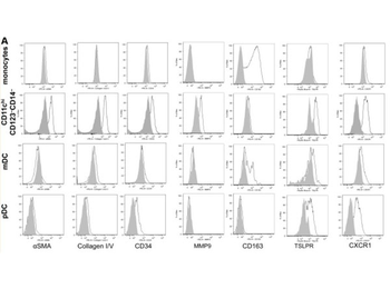
Flow cytometry of Anti-Collagen Type I Antibody Biotin Conjugated. Expanded CD11chiCD123–CD14– cells are fibrocytes that mediate angiogenesis. (A) Using the same gating strategy as shown in Figure 1A, CD11chiCD123–CD14– cells from a representative subject sample were analyzed for cell surface phenotype. The shaded areas represent background fluorescence on the designated population as indicated by FMO controls. This is representative of more than 10 experiments.
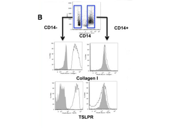
Flow Cytometry of Anti-Collagen Type I Antibody Biotin Conjugated. IL-4 induces monocytes to differentiate into CD14– fibrocytes that are readily distinguished from CD14 + macrophages in the same culture. (B) Cell surface phenotype of IL-4–differentiated adherent cells identifies 2 subsets based on CD14 expression, which further shows differential expression of collagen and TSLPR. FMO controls on gated CD14 + vs CD14– populations are shown by shaded gray histograms. This is representative of more than 5 experiments from 5 separate healthy donors.
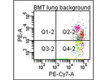
Flow Cytometry of Anti-Collagen Type I Biotin Conjugated Antibody (orb345868). Cells: mouse lung. Stimulation: none. Primary antibody: biotin conjugated anti-collagen type I antibody. Secondary antibody: PE-conjugated CD45 and PE-conjugated anti-collagen type I secondary antibody.
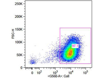
Flow Cytometry of Rabbit Anti-Collagen 1 Antibody. Cells: primary adult human dermal fibroblast cells. Stimulation: none. Primary antibody: Biotin-Conjugated Collagen 1 antibody (orb345868) at 5 µg/ml for 45 min at 4°C. Secondary antibody: Rabbit Streptavidin, R-PE antibody at 1:500 for 15 min at RT.
Quick Database Links
UniProt Details
− No UniProt data available
NCBI Reference Sequences
−Associated Accession Numbers
Curated reference sequences for the gene transcript and protein product| Protein | NP_000079.2 |
|---|
Documents Download
Datasheet
Product Information
Request a Document
Protocol Information
WB
Western Blot (IB, immunoblot)
IHC
Immunohistochemistry
FC
Flow Cytometry
ELISA
Enzyme-linked Immunosorbent Assay (EIA)
IP
Immunoprecipitation
DOT
Dot Blot
COL1A1 Antibody (Biotin) (orb345868)

- 0.0
Based on 0 reviews
Participating in our Biorbyt product reviews program enables you to support fellow scientists by sharing your firsthand experience with our products.
Login to Submit a ReviewAvailable Sizes
Select a size below
Choose Conjugation or Carrier Free Version
Free Secondary Antibody (20 ul)0/0
Please add an antibody product to your cart first.