You have no items in your shopping cart.
Featured
Description
Research Area
Cell Biology
Images & Validation
−Item 1 of 3
| Tested Applications | ICC, IF, WB |
|---|---|
| Dilution Range | WB (1:1000), ICC/IF (1:100) |
| Reactivity | Human |
| Application Notes |
Key Properties
−| Host | Mouse |
|---|---|
| Clonality | Monoclonal |
| Isotype | IgG1 Kappa |
| Clone No. | 1C4-1A6 |
| Immunogen | Human HSP47, full length |
| Target | HSP47 |
| Molecular Weight | 47kDa |
| Purification | Protein G Purified |
| Conjugation | Unconjugated |
Storage & Handling
−| Storage | Maintain refrigerated at 2-8°C for up to 2 weeks. For long term storage store at -20°C in small aliquots to prevent freeze-thaw cycles. |
|---|---|
| Buffer/Preservatives | PBS pH 7.4, 50% glycerol, 0.09% sodium azide. Storage buffer changes when conjugated. |
| Concentration | 1 mg/ml |
| Expiration Date | 12 months from date of receipt. |
| Disclaimer | For research use only |
Alternative Names
−SERPINH1, HSP47, Heat shock protein 47, Serpin H1, Colligin, Collagen-binding protein, Gp46, Serine protease inhibitor J6, 47 kDa heat shock protein, serine proteinase inhibitor, cysteine proteinase inhibitor, collagen binding protein
Similar Products
−- Item 1 of 11
HSP47 Rabbit Polyclonal Antibody [orb2563448]
IF, IHC-Fr, IHC-P
Mouse, Rat
Human, Mouse, Rat
Rabbit
Polyclonal
Unconjugated
50 μl, 100 μl, 200 μl - Item 1 of 7
Hsp47/SERPINH1 Rabbit Polyclonal Antibody [orb234371]
FC, ICC, IHC, WB
Human, Mouse, Rat
Rabbit
Polyclonal
Unconjugated
100 μg - Item 1 of 1
- Item 1 of 3
SERPINH1 Antibody (Center) [orb1929322]
FC, IHC-P, WB
Mouse, Rat
Human
Rabbit
Polyclonal
Unconjugated
100 μl, 50 μl - Item 1 of 3
SERPINH1 Antibody (C-term) [orb1929323]
FC, IHC-P, WB
Mouse, Rat
Human
Rabbit
Polyclonal
Unconjugated
50 μl, 100 μl
Quality Guarantee
Explore bioreagents carefree to elevate your research. All our products are rigorously tested for performance. If a product does not perform as described on its datasheet, our scientific support team will provide expert troubleshooting, a prompt replacement, or a refund. For full details, please see our Terms & Conditions and Buying Guide. Contact us at [email protected].
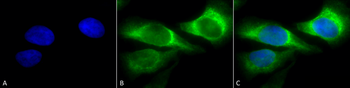
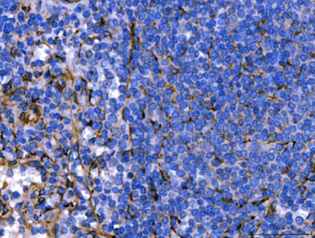
Immunocytochemistry/Immunofluorescence analysis using Mouse Anti-Hsp47 Monoclonal Antibody, Clone 1C4-1A6. Tissue: Heat Shocked cervical cancer cells (HeLa). Species: Human. Fixation: 2% Formaldehyde for 20 min at RT. Primary Antibody: Mouse Anti-Hsp47 Monoclonal Antibody at 1:100 for 12 hours at 4°C. Secondary Antibody: FITC Goat Anti-Mouse (green) at 1:200 for 2 hours at RT. Counterstain: DAPI (blue) nuclear stain at 1:40000 for 2 hours at RT. Localization: Endoplasmic reticulum lumen. Cytoplasm. Magnification: 100x. (A) DAPI (blue) nuclear stain. (B) Anti-Hsp47 Antibody. (C) Composite. Heat Shocked at 42°C for 1h.
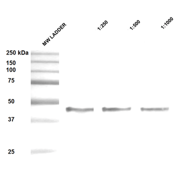
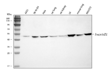
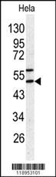
Western Blot analysis of Human Epithelial cell (A431) lysates showing detection of ~47 kDa Hsp47 protein using Mouse Anti-Hsp47 Monoclonal Antibody, Clone 1C4-1A6. Lane 1: MW ladder. Lane 2: Anti-Hsp47 (1:250). Lane 3: Anti-Hsp47 (1:500). Lane 4: Anti-Hsp47 (1:1000). Load: 20 μg. Block: 5% milk + TBST for 1 hour at RT. Primary Antibody: Mouse Anti-Hsp47 Monoclonal Antibody at 1:250 - 1:1000 for 1 hour at RT. Secondary Antibody: HRP Goat Anti-Mouse at 1:50 for 1 hour at RT. Color Development: TMB solution for 10 min at RT. Predicted/Observed Size: ~47 kDa.
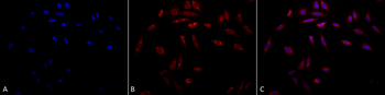
Immunocytochemistry/Immunofluorescence analysis using Mouse Anti-Hsp47 Monoclonal Antibody, Clone 1C4-1A6. Tissue: Heat Shocked cervical cancer cells (HeLa). Species: Human. Fixation: 2% Formaldehyde for 20 min at RT. Primary Antibody: Mouse Anti-Hsp47 Monoclonal Antibody at 1:100 for 12 hours at 4°C. Secondary Antibody: APC Goat Anti-Mouse (red) at 1:200 for 2 hours at RT. Counterstain: DAPI (blue) nuclear stain at 1:40000 for 2 hours at RT. Localization: Endoplasmic reticulum lumen. Cytoplasm. Magnification: 20x. (A) DAPI (blue) nuclear stain. (B) Anti-Hsp47 Antibody. (C) Composite. Heat Shocked at 42°C for 1h.
Quick Database Links
UniProt Details
− No UniProt data available
NCBI Gene Details
− No NCBI Gene data available
NCBI Reference Sequences
−Associated Accession Numbers
Curated reference sequences for the gene transcript and protein product| Protein | NP_001193943 |
|---|
Documents Download
Datasheet
Product Information
Request a Document
Protocol Information
WB
Western Blot (IB, immunoblot)
IF
Immunofluorescence
ICC
Immunocytochemistry
HSP47 Antibody (orb99078)

- 0.0
Based on 0 reviews
Participating in our Biorbyt product reviews program enables you to support fellow scientists by sharing your firsthand experience with our products.
Login to Submit a ReviewAvailable Sizes
Select a size below
Free Secondary Antibody (20 ul)0/0
Please add an antibody product to your cart first.