You have no items in your shopping cart.
F11R Antibody
SKU: orb420322
Description
Research Area
Immunology & Inflammation
Images & Validation
−Item 1 of 7
| Tested Applications | ELISA, IF, WB |
|---|---|
| Dilution Range | ELISA: 5 ug/ml, WB: 1 ug/ml |
| Reactivity | Human |
| Application Notes |
Key Properties
−| Antibody Type | Primary Antibody |
|---|---|
| Host | Rabbit |
| Clonality | Polyclonal |
| Isotype | IgG |
| Immunogen | Affinity purified Anti-JAM A pY280 antibody was prepared from whole rabbit serum produced by repeated immunizations with a synthetic peptide corresponding to the c-term and phosphorylated at the tyrosine 280 position of Human JAM A protein. |
| Target | F11R |
| Purity | Anti-JAM A pY280 is directed against human JAM A phosphorylated at the tyrosine 280 position. This product is an affinity purified antibody produced by immunoaffinity chromatography using phospho peptide coupled to agarose beads followed by solid phase adsorption(s) against non-phospho peptide to remove any unwanted reactivities. A BLAST analysis was used to suggest reactivity with this protein from human and feline based on 100% homology for the immunogen sequence. |
| Conjugation | Unconjugated |
Storage & Handling
−| Storage | Store vial at -20° C prior to opening. Aliquot contents and freeze at -20° C or below for extended storage. Avoid cycles of freezing and thawing. Centrifuge product if not completely clear after standing at room temperature. This product is stable for several weeks at 4° C as an undiluted liquid. Dilute only prior to immediate use. |
|---|---|
| Form/Appearance | Liquid (sterile filtered) |
| Buffer/Preservatives | Preservative: 0.01% (w/v) Sodium Azide. Stabilizer: None; Buffer: 0.02 M Potassium Phosphate, 0.15 M Sodium Chloride, pH 7.2 |
| Concentration | 1.02mg/mL |
| Expiration Date | 12 months from date of receipt. |
| Dry Ice Shipping | Please note: This product requires shipment on dry ice. A dry ice surcharge will apply. |
| Disclaimer | For research use only |
Alternative Names
−rabbit anti-JAM A pY280 antibody, JAM-A, Junctional adhesion molecule A, JAM-1, Junctional adhesion molecule 1, Platelet F11 receptor, Platelet adhesion molecule 1, PAM-1, CD321, JAM1, JCAM, JAM 1, JAMA
Similar Products
−- Item 1 of 7
- Item 1 of 5
JAM-A/F11R/JAM Rabbit Polyclonal Antibody [orb1147774]
ELISA, FC, IHC, WB
Human
Rabbit
Polyclonal
Unconjugated
100 μg - Item 1 of 4
- Item 1 of 1
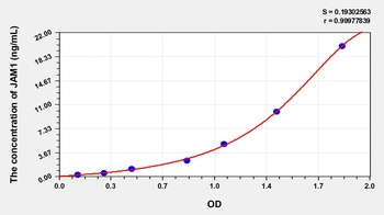
Human Junctional Adhesion Molecule 1 (JAM1) ELISA Kit [orb777786]
Human
0.32-20 ng/mL
0.133 ng/mL
48 T, 96 T - Item 1 of 3


Quality Guarantee
Explore bioreagents carefree to elevate your research. All our products are rigorously tested for performance. If a product does not perform as described on its datasheet, our scientific support team will provide expert troubleshooting, a prompt replacement, or a refund. For full details, please see our Terms & Conditions and Buying Guide. Contact us at [email protected].

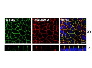
Confocal Immunofluorescence Microscopy of Rabbit Anti-JAM-A pY280 antibody in polarized epithelial cells. Tissue: T84 cells were grown on Transwell filters until confluent. Treatment: pervanadate. Fixation: 4% PFA. Permeabilization: 1% SDS. Costained Green: Anti-Phospho JAM-A Y280 Antibody, FITC conjugated secondary; Red: Anti-Total JAM-A, Alexa-conjugated secondary antibodies. Localization: tight junctions, seen in Confocal Z-stacks. Scale bar: 10 µm.
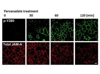
Confocal Immunofluorescence Microscopy of Rabbit Anti-JAM-A pY280 antibody of confluent (intestinal epithelial cells) IECs. Tissue: SK CO-15 cells. Treatment: pervanadate at time points 0, 30, 60, 120 mins. Fixation: 4% PFA. Permeabilization: 1% SDS. Costained Green: Anti-Phospho JAM-A Y280 Antibody, FITC conjugated secondary; Red: Anti-Total JAM-A, Alexa-conjugated secondary antibodies. Results: pervanadate treatment led to a time-dependent increase in phosphorylation of JAM-A Y280 that correlated with decreased localization of JAM-A at cell–cell contacts. Scale bar: 10 µm.
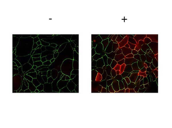
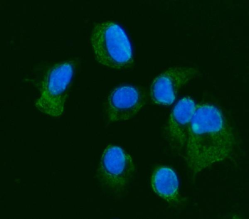
Immunofluorescence Microscopy of Rabbit anti-JAMA pY280 antibody. Tissue: T84 cells (untreated/treated). Fixation: 0.5% PFA. Antigen retrieval: not required. Primary antibody: JAMA pY280 antibody at 2 µg/ml for 1 hr at RT. Secondary antibody: Fluorescein rabbit secondary antibody at 1:10000 for 45 min at RT. Localization: JAMA pY280 is along the cell membrane and cell junction. Staining: JAMA pY280 as red fluorescent signal.
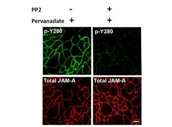
Immunofluorescence Microscopy of Rabbit Anti-JAM-A pY280 antibody. Tissue: T84 cells. Pretreatment: PP2. Treatment: Pervanadate. Fixation: 4% PFA. Permeabilization: 1% SDS. Costained Green: Anti-Phospho JAM-A Y280 Antibody, FITC conjugated secondary; Red: Anti-Total JAM-A, Alexa-conjugated secondary antibodies. Results: The Src family kinase inhibitor PP2 inhibits pervanadate-dependent phosphorylation of JAM-A Y280, as they reported to modulate tyrosine phosphorylation of junctional proteins and influence epithelial barrier function.
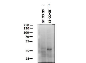
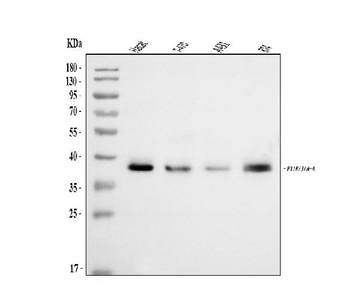
Western Blot of Rabbit anti-JAM A pY280 antibody. Lane 1: SK-CO-15 negative control. Lane 2: SK-CO-15 pervanadate treated positive control. Load: 10 µg per lane. Primary antibody: JAM A pY280 antibody at 1 ug/ml for overnight at 4°C. Secondary antibody: Peroxidase rabbit secondary antibody at 1:40000 for 30 min at RT. Block: orb348637 for 30 minutes at RT. Predicted/Observed size: ~ 32.5 kDa. JCYIA
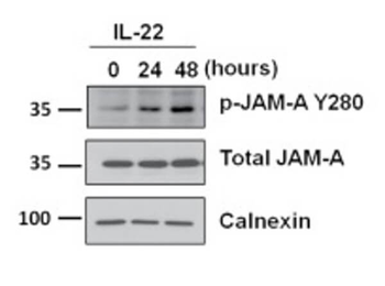
Western Blot of Rabbit Anti-JAM-A pY280 antibody with IL-22. Lysates: T84 cells. Treatment: hu rec. IL-22 at time points 0, 24, 48 hrs. Primary antibodies: JAM-A pY280, total JAM-A, or Calnexin. Calnexin was used as a loading control. Secondary antibody: horseradish peroxidase secondary antibody. Results: Exposure of IECs to other cytokines (IL-17A, IL-22, TNFα, or IFNγ) results in tyrosine phosphorylation of JAM-A at Y280 and a leaky barrier.
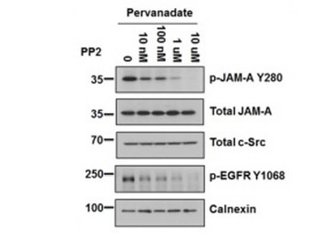
Western Blot of Rabbit Anti-JAM-A pY280 antibody with PP2. Lysates: T84 cells. Treatments: Pervanadate; PP2 at 0, 10 nM, 100 nM, 1 µm, 10 µm. Primary antibodies: p-JAM-A Y280, total JAM-A, total c-Src, p-EGFR Y1068, or Calnexin. p-EGFR Y1068 was used as a positive control for PP2. Calnexin was used as a loading control. Secondary antibody: horseradish peroxidase secondary antibody. Results: PP2 dose-dependent decrease in tyrosine phosphorylation of JAM-A Y280 following pervanadate treatment.
Quick Database Links
UniProt Details
− No UniProt data available
NCBI Reference Sequences
−Associated Accession Numbers
Curated reference sequences for the gene transcript and protein product| Protein | NP_058642.1 |
|---|
Documents Download
Datasheet
Product Information
Request a Document
Protocol Information
WB
Western Blot (IB, immunoblot)
IF
Immunofluorescence
ELISA
Enzyme-linked Immunosorbent Assay (EIA)
F11R Antibody (orb420322)

- 0.0
Based on 0 reviews
Participating in our Biorbyt product reviews program enables you to support fellow scientists by sharing your firsthand experience with our products.
Login to Submit a ReviewAvailable Sizes
Select a size below