You have no items in your shopping cart.
Description
Research Area
Cell Biology
Images & Validation
−Item 1 of 4
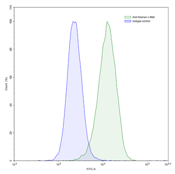
| Tested Applications | IF, IHC-P, WB |
|---|---|
| Dilution Range | IHC-P - 1:50-100, WB - 1:8000, IF - 1:100 |
| Reactivity | Human, Mouse |
Key Properties
−| Host | Mouse |
|---|---|
| Clonality | Monoclonal |
| Isotype | Mouse IgG1 |
| Clone No. | B6CV66 |
| Immunogen | This monoclonal antibody is generated from mice immunized with purified recombinant protein encoding the catalytic domain of human Met. Antigen Region: Unknown. |
| Target | MET |
| Molecular Weight | 155541 Da |
| Conjugation | Unconjugated |
Storage & Handling
−| Storage | Maintain refrigerated at 2-8°C for up to 2 weeks. For long term storage store at -20°C in small aliquots to prevent freeze-thaw cycles |
|---|---|
| Form/Appearance | Purified monoclonal antibody supplied in PBS with 0.09% (W/V) sodium azide. This antibody is purified through a protein G column, followed by dialysis against PBS. |
| Expiration Date | 12 months from date of receipt. |
| Disclaimer | For research use only |
Alternative Names
−Hepatocyte growth factor receptor, HGF receptor, HGF/SF receptor, Proto-oncogene c-Met, Scatter factor receptor, SF receptor, Tyrosine-protein kinase Met, MET
Similar Products
−- Item 1 of 3
- Item 1 of 3
- Item 1 of 3
- Item 1 of 3
MET/c-Met/HGFR Human Monoclonal Antibody [orb2975373]
FC
Human
Human
Monoclonal
Unconjugated
50 μg, 100 μg - Item 1 of 3
MET/c-Met/HGFR Human Monoclonal Antibody [orb2975374]
FC
Human
Human
Monoclonal
Unconjugated
50 μg, 100 μg


Quality Guarantee
Explore bioreagents carefree to elevate your research. All our products are rigorously tested for performance. If a product does not perform as described on its datasheet, our scientific support team will provide expert troubleshooting, a prompt replacement, or a refund. For full details, please see our Terms & Conditions and Buying Guide. Contact us at [email protected].
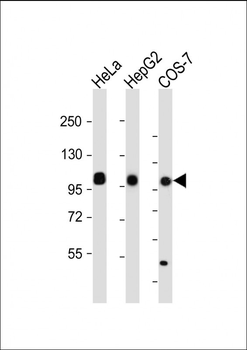
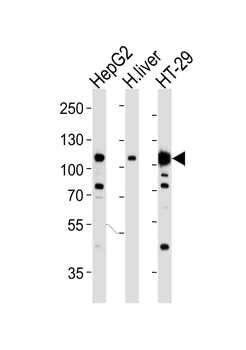
All lanes: Anti-MET/HGFR Antibody at dilution. Lane 1: HeLa whole cell lysate. Lane 2: HepG2 whole cell lysate. Lane 3: COS-7 whole cell lysate.Lysates/proteins at 20 µg per lane. Secondary Goat Anti-mouse IgG, (H+L), Peroxidase conjugated at 1/10000 dilution. Predicted band size: 156 kDa. Blocking/Dilution buffer: 5% NFDM/TBST.
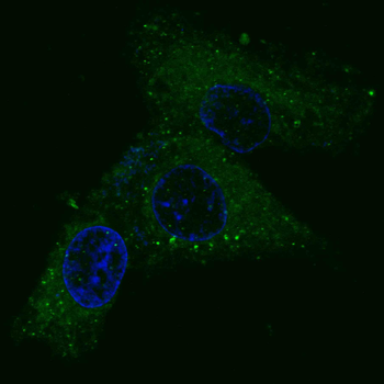
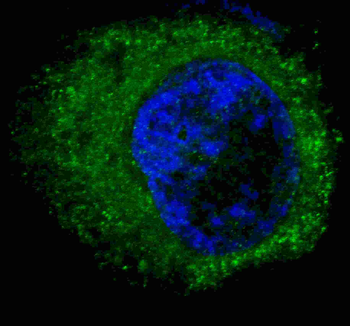
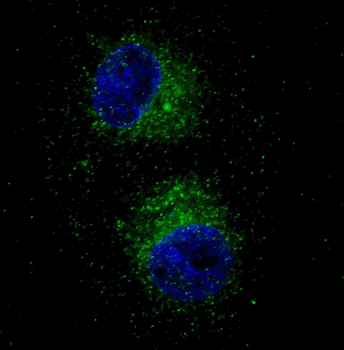
Fluorescent confocal image of HepG2 cells stained with MET/HGFR Antibody. HepG2 cells were fixed with 4% PFA (20 min), permeabilized with Triton X-100 (0.2%, 30 min). Cells were then incubated with MET/HGFR primary antibody (1:100, 2 h at room temperature). For secondary antibody, Alexa Fluor 488 conjugated donkey anti-mouse antibody (green) was used (1:1000, 1h). Nuclei were counterstained with Hoechst 33342 (blue) (10 μg/ml, 5 min).
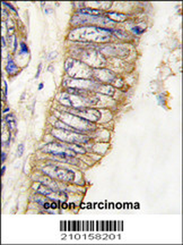
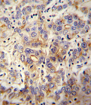
Formalin-fixed and paraffin-embedded human colon carcinoma tissue reacted with MET/HGFR Antibody, which was peroxidase-conjugated to the secondary antibody, followed by DAB staining. This data demonstrates the use of this antibody for immunohistochemistry; clinical relevance has not been evaluated.
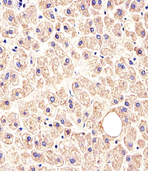
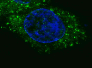
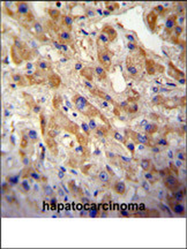
Immunohistochemical analysis of paraffin-embedded H.liver section using MET/HGFR Antibody. diluted at 1:25 dilution. A peroxidase-conjugated goat anti-mouse IgG at 1:400 dilution was used as the secondary antibody, followed by DAB staining.
Quick Database Links
UniProt Details
− No UniProt data available
NCBI Reference Sequences
−Associated Accession Numbers
Curated reference sequences for the gene transcript and protein product| Protein | NP_001120972.1, NP_000236.2 |
|---|
Documents Download
Datasheet
Product Information
Request a Document
Protocol Information
WB
Western Blot (IB, immunoblot)
IHC-P
Immunohistochemistry Paraffin
IF
Immunofluorescence
MET/HGFR Antibody (orb1939458)

- 0.0
Based on 0 reviews
Participating in our Biorbyt product reviews program enables you to support fellow scientists by sharing your firsthand experience with our products.
Login to Submit a ReviewAvailable Sizes
Select a size below
Choose Conjugation or Carrier Free Version
Free Secondary Antibody (20 ul)0/0
Please add an antibody product to your cart first.