You have no items in your shopping cart.
Description
Research Area
Immunology & Inflammation
Images & Validation
−Item 1 of 6
| Tested Applications | FC, ICC, IHC, WB |
|---|---|
| Dilution Range | WB - 1:1000; IHC - 1:100 - 1:500. Epitope retrieval with citrate buffer pH 6.0 is recommended for FFPE tissue sections.; ICC - 1:100 - 1:500. Epitope retrieval with citrate buffer pH 6.0 is recommended for FFPE cell sections.; IHC-IF - 1:100 to 1:500. Epitope retrieval with citrate buffer pH6.0 is recommended for FFPE cell sections. |
| Reactivity | Human |
| Application Notes |
Key Properties
−| Antibody Type | Primary Antibody |
|---|---|
| Host | Mouse |
| Clonality | Monoclonal |
| Isotype | IgG2b |
| Clone No. | LN3 |
| Immunogen | Activated human peripheral blood mononuclear cells |
| Target | HLA-DR |
| Purification | Purified |
| Conjugation | Unconjugated |
Storage & Handling
−| Storage | 2 - 8°C |
|---|---|
| Form/Appearance | Liquid |
| Buffer/Preservatives | Phosphate Buffered Saline (PBS) pH 8.2 with 0.1% rAlbumin and 0.09% Sodium Azide |
| Concentration | 50 µg/ml |
| Expiration Date | 12 months from date of receipt. |
| Disclaimer | For research use only |
Alternative Names
−histocompatibility antigen HLA-DR alpha; HLA class II histocompatibility antigen, DR alpha chain; HLA-DRA1; MHC class II antigen DRA
Similar Products
−- Item 1 of 7
HLA-DR/HLA-DRA/HLA Mouse Monoclonal Antibody [orb1152388]
FC, IHC, WB
Human
Mouse
Monoclonal
Unconjugated
100 μg - Item 1 of 5
- Item 1 of 6
HLA-DR/HLA-DRA/HLA Mouse Monoclonal Antibody [orb1152389]
FC, IHC, WB
Human
Mouse
Monoclonal
Unconjugated
100 μg - Item 1 of 4
HLA-DRA Antibody [orb1410572]
FC, IF, IHC, WB
Human
Mouse
Monoclonal
Unconjugated
20 μg, 100 μg, 100 μg (without BSA and Azide) - Item 1 of 3


![Anti-HLA class II [F3.3]](/images/pub/media/catalog/product/NewWebsite/35/orb411559_1.png)
![Anti-HLA class II [F3.3]](/images/pub/media/catalog/product/NewWebsite/35/orb411559_2.png)
![Anti-HLA class II [F3.3]](/images/pub/media/catalog/product/NewWebsite/35/orb411559_3.png)
Quality Guarantee
Explore bioreagents carefree to elevate your research. All our products are rigorously tested for performance. If a product does not perform as described on its datasheet, our scientific support team will provide expert troubleshooting, a prompt replacement, or a refund. For full details, please see our Terms & Conditions and Buying Guide. Contact us at [email protected].

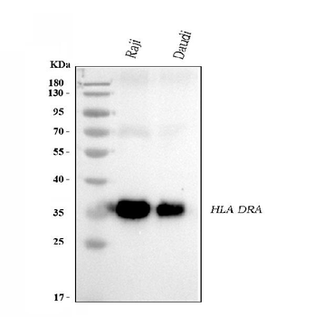
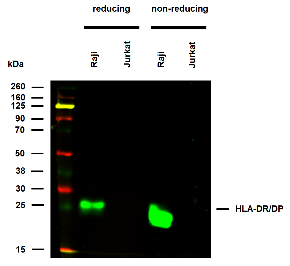
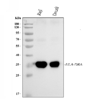
Detection of human HLA-DR by western blot. Samples: Whole cell lysate (50 µg) from KG-1, SNB-75, Raji, Ramos, and SR (10 µg) cells prepared using NETN lysis buffer. Antibody: Mouse anti-HLA-DR monoclonal antibody [LN3] (orb1520116) used at 1:1000. Secondary: HRP-conjugated goat anti-mouse IgG. Detection: Chemiluminescence with an exposure time of 3 minutes. Lower Panel: Rabbit anti-Actin recombinant monoclonal antibody.

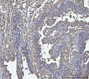
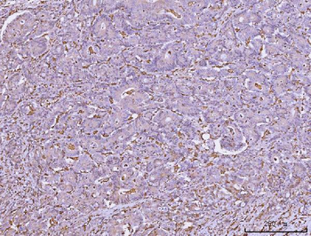
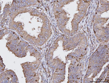
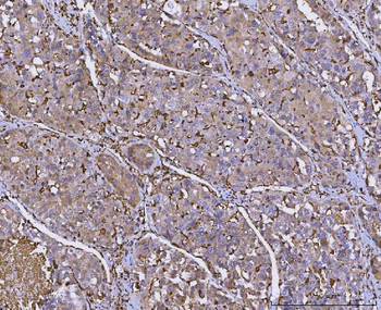
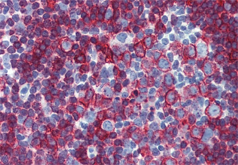
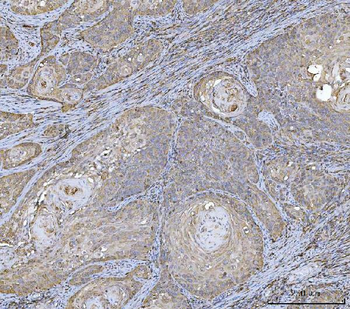
Detection of human HLA-DR by immunohistochemistry. Sample: FFPE section of tonsil. Antibody: Mouse anti-HLA-DR monoclonal antibody [LN3]. Secondary: HRP-conjugated goat anti-mouse IgG.

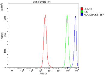
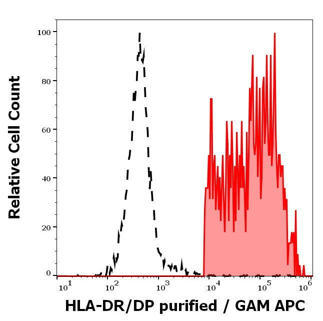
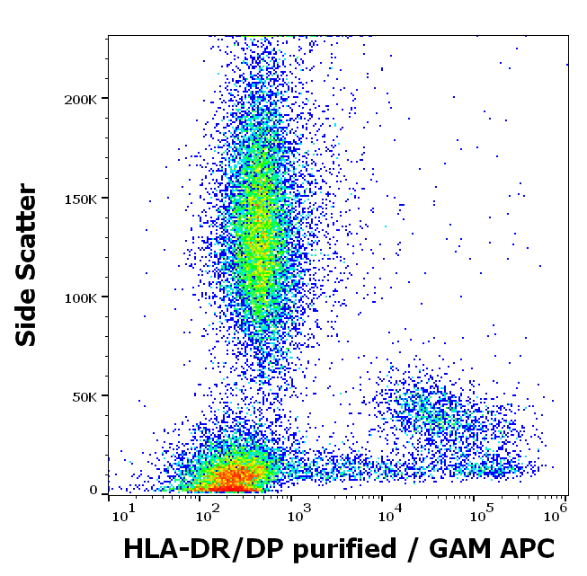
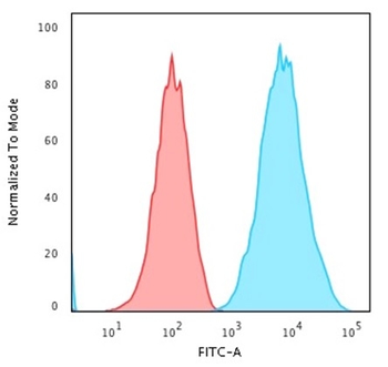
Detection of human HLA-DR (shaded) in Daudi cells by flow cytometry. Antibody: Mouse anti-HLA-DR monoclonal antibody [LN3] (orb1520116) or isotype control (unshaded). Secondary: DyLight® 488-conjugated goat anti-mouse IgG.

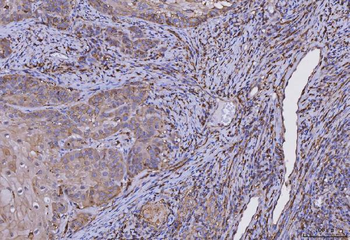
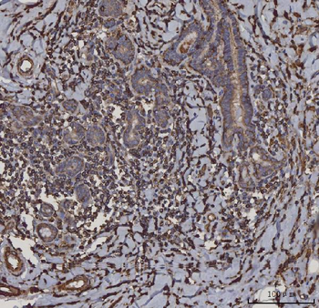
Detection of human HLA-DR by immunocytochemistry. Sample: FFPE section of SR cells. Antibody: Mouse anti-HLA-DR monoclonal antibody [LN3] (orb1520116). Secondary: HRP-conjugated goat anti-mouse IgG.

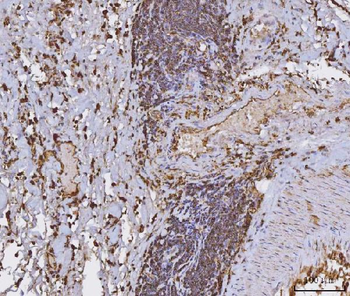
Detection of human HLA-DR by immunhistochemistry. Sample: FFPE section of human tonsil. Antibody: Mouse monoclonal anti-HLA-DR antibody [LN3] (orb1520116) used at 1:100. Secondary: DyLight® 594-conjugated goat anti-mouse IgG.

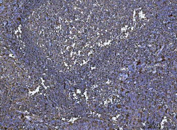
Detection of human HLA-DR (red) by immunohistochemistry. Sample: FFPE section of human tonsil. Antibody: Mouse anti-HLA-DR monoclonal antibody [LN3] (orb1520116) used at 1:250. Secondary: HRP-conjugated goat anti-mouse IgG. Substrate: Opal™. Counterstain: DAPI (blue).
Quick Database Links
UniProt Details
− No UniProt data available
NCBI Reference Sequences
−Associated Accession Numbers
Curated reference sequences for the gene transcript and protein product| Protein | NP_061984.2 |
|---|
Documents Download
Datasheet
Product Information
Request a Document
Protocol Information
WB
Western Blot (IB, immunoblot)
IHC
Immunohistochemistry
FC
Flow Cytometry
ICC
Immunocytochemistry
Mouse HLA-DR Monoclonal Antibody (orb1520116)

- 0.0
Based on 0 reviews
Participating in our Biorbyt product reviews program enables you to support fellow scientists by sharing your firsthand experience with our products.
Login to Submit a ReviewAvailable Sizes
Select a size below
Choose Conjugation or Carrier Free Version
Free Secondary Antibody (20 ul)0/0
Please add an antibody product to your cart first.