You have no items in your shopping cart.
Description
Research Area
Cell Biology
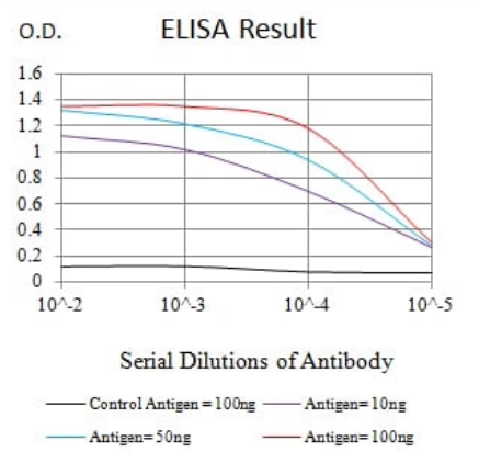
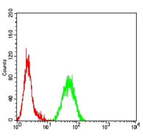
Images & Validation
−


Item 1 of 5
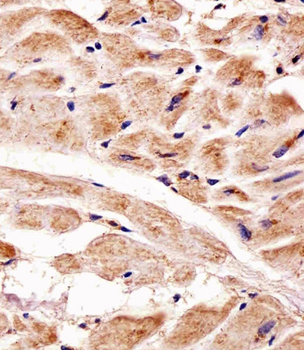
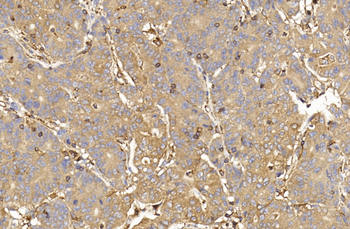
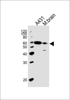
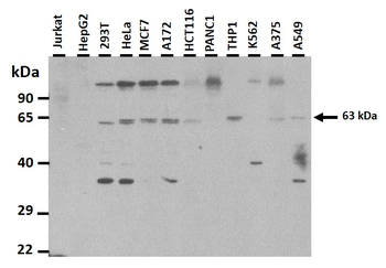
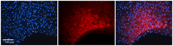
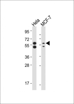
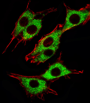
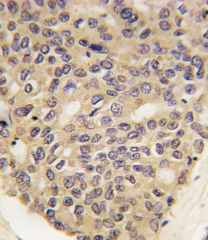
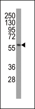
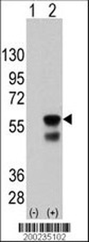
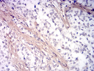
| Tested Applications | IF, IHC-P, WB |
|---|---|
| Dilution Range | IF: 1:25, WB: 1:1000, IHC-P: 1:25, IHC-P: 1:25, IHC-P: 1:25 |
| Reactivity | Human, Mouse, Rat |
Key Properties
−| Antibody Type | Primary Antibody |
|---|---|
| Host | Mouse |
| Clonality | Monoclonal |
| Isotype | IgG1 |
| Immunogen | Recombinant PINK1 protein was used to produced this monoclonal antibody. Antigen Region: Unknown. |
| Target | PINK1 |
| Molecular Weight | 62769 Da |
| Conjugation | Unconjugated |
Storage & Handling
−| Storage | Maintain refrigerated at 2-8°C for up to 2 weeks. For long term storage store at -20°C in small aliquots to prevent freeze-thaw cycles |
|---|---|
| Form/Appearance | Purified monoclonal antibody supplied in PBS with 0.09% (W/V) sodium azide. This antibody is purified through a protein G column, followed by dialysis against PBS. |
| Expiration Date | 12 months from date of receipt. |
| Disclaimer | For research use only |
Alternative Names
−Serine/threonine-protein kinase PINK1, mitochondrial, BRPK, PTEN-induced putative kinase protein 1, PINK1
Similar Products
−- Item 1 of 4
PINK1 Rabbit Polyclonal Antibody [orb331223]
IHC, WB
Bovine, Equine, Guinea pig, Mouse, Rabbit, Rat
Human
Rabbit
Polyclonal
Unconjugated
100 μl - Item 1 of 4
- Item 1 of 4
PINK1 Antibody (Ascites) [orb1440457]
IHC-P, WB
Human, Mouse
Mouse
Monoclonal
Unconjugated
50 μl, 100 μl - Item 1 of 1
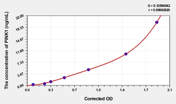
Human Serine/threonine-protein kinase PINK1, Mitochondrial (PINK1) ELISA Kit [orb1736595]
Human
0.32-20 ng/mL
0.14 ng/mL
48 T, 96 T - Item 1 of 5


Quality Guarantee
Explore bioreagents carefree to elevate your research. All our products are rigorously tested for performance. If a product does not perform as described on its datasheet, our scientific support team will provide expert troubleshooting, a prompt replacement, or a refund. For full details, please see our Terms & Conditions and Buying Guide. Contact us at [email protected].
Quick Database Links
UniProt Details
− No UniProt data available
NCBI Reference Sequences
−Associated Accession Numbers
Curated reference sequences for the gene transcript and protein product| Protein | NP_115785.1 |
|---|
Protocol Information
WB
Western Blot (IB, immunoblot)
IHC-P
Immunohistochemistry Paraffin
IF
Immunofluorescence