You have no items in your shopping cart.
Description
Research Area
Cardiovascular Research, Cell Biology, Infectious Disease & Virology, Metabolism Research
Images & Validation
−Item 1 of 4
| Tested Applications | FC, IHC-P, WB |
|---|---|
| Dilution Range | WB - 1:1000, IHC-P - 1:50-100, FC - 1:10-50 |
| Reactivity | Human |
| Predicted Reactivity | Mouse, Porcine, Rat |
Key Properties
−| Antibody Type | Primary Antibody |
|---|---|
| Host | Rabbit |
| Clonality | Polyclonal |
| Isotype | Rabbit IgG |
| Immunogen | This SUMO1 antibody is generated from rabbits immunized with a KLH conjugated synthetic peptide between 55-86 amino acids from the C-terminal region of human SUMO1. Antigen Region: 55-86 aa. |
| Target | SUMO1 |
| Molecular Weight | 11557 Da |
| Conjugation | Unconjugated |
Storage & Handling
−| Storage | Maintain refrigerated at 2-8°C for up to 2 weeks. For long term storage store at -20°C in small aliquots to prevent freeze-thaw cycles |
|---|---|
| Form/Appearance | Purified polyclonal antibody supplied in PBS with 0.09% (W/V) sodium azide. This antibody is prepared by Saturated Ammonium Sulfate (SAS) precipitation followed by dialysis against PBS. |
| Expiration Date | 12 months from date of receipt. |
| Disclaimer | For research use only |
Alternative Names
−Small ubiquitin-related modifier 1, SUMO-1, GAP-modifying protein 1, GMP1, SMT3 homolog 3, Sentrin, Ubiquitin-homology domain protein PIC1, Ubiquitin-like protein SMT3C, Smt3C, Ubiquitin-like protein UBL1, SUMO1, SMT3C, SMT3H3, UBL1
Similar Products
−- Item 1 of 1
SUMO1 Antibody (C-term D86) [orb1939632]
WB
Mouse, Other, Porcine, Rat, Zebrafish
Human
Rabbit
Polyclonal
Unconjugated
50 μl, 100 μl
Quality Guarantee
Explore bioreagents carefree to elevate your research. All our products are rigorously tested for performance. If a product does not perform as described on its datasheet, our scientific support team will provide expert troubleshooting, a prompt replacement, or a refund. For full details, please see our Terms & Conditions and Buying Guide. Contact us at [email protected].
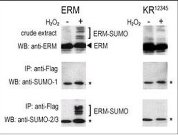
COS-7 cells were transfected for 24 hrs with a plasmid expressing FLAG-ERM (left panels) or FLAG-ERM KR12345 (right panels). Untreated (-) and H2O2-treated (+) cells were collected for immunoblot analysis. Top panels: cell lysates probed by western blot (WB) with an anti-ERM Antibody. Center panels: cell lysates immunoprecipitated (IP) with an anti-FLAG antibody followed by WB with SUMO-1 Antibody. Bottom panels: cell lysates immunoprecipitated with an anti-FLAG antibody followed by WB with SUMO-2/3 Antibody. (*) represents immunoprecipitated ERM-like forms recognized by anti-SUMO antibodies.
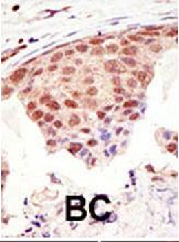
Formalin-fixed and paraffin-embedded human cancer tissue reacted with the primary antibody, which was peroxidase-conjugated to the secondary antibody, followed by DAB staining. This data demonstrates the use of this antibody for immunohistochemistry; clinical relevance has not been evaluated. BC = breast carcinoma; HC = hepatocarcinoma.
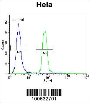
SUMO1 Antibody (C-term) flow cytometric analysis of Hela cells (right histogram) compared to a negative control cell (left histogram). FITC-conjugated goat-anti-rabbit secondary antibodies were used for the analysis.
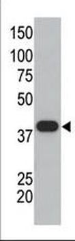
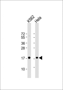
The anti-SUMO1 polyclonal antibody is used in Western blot to detect GST-SUMO1 fusion protein.
Quick Database Links
UniProt Details
− No UniProt data available
NCBI Reference Sequences
−Associated Accession Numbers
Curated reference sequences for the gene transcript and protein product| Protein | NP_003343.1, NP_001005782.1, NP_001005781.1 |
|---|
Documents Download
Datasheet
Product Information
Request a Document
Protocol Information
WB
Western Blot (IB, immunoblot)
IHC-P
Immunohistochemistry Paraffin
FC
Flow Cytometry
SUMO1 Antibody (C-term) (orb1937645)

- 0.0
Based on 0 reviews
Participating in our Biorbyt product reviews program enables you to support fellow scientists by sharing your firsthand experience with our products.
Login to Submit a ReviewAvailable Sizes
Select a size below
Choose Conjugation or Carrier Free Version
Free Secondary Antibody (20 ul)0/0
Please add an antibody product to your cart first.