
You have no items in your shopping cart.
Featured
Description
Research Area
Cell Biology, Metabolism Research, Signal Transduction
Images & Validation
−Item 1 of 7
| Tested Applications | FC, IHC-P, WB |
|---|---|
| Dilution Range | WB - 1:1000, IHC-P-Leica - 1:500, IHC-P - 1:100, FC - 1:25 |
| Reactivity | Human, Mouse, Rat |
| Predicted Reactivity | Porcine, Rabbit |
Key Properties
−| Antibody Type | Primary Antibody |
|---|---|
| Host | Rabbit |
| Clonality | Polyclonal |
| Isotype | Rabbit IgG |
| Immunogen | Synthetic Peptide |
| Target | VDAC3 |
| Molecular Weight | 30659 |
| Conjugation | Unconjugated |
Storage & Handling
−| Storage | Maintain refrigerated at 2-8°C for up to 2 weeks. For long term storage store at -20°C in small aliquots to prevent freeze-thaw cycles |
|---|---|
| Form/Appearance | Purified polyclonal antibody supplied in PBS with 0.09% (W/V) sodium azide. This antibody is purified through a protein A column, followed by peptide affinity purification. |
| Expiration Date | 12 months from date of receipt. |
| Disclaimer | For research use only |
Alternative Names
−Anti-Voltage-dependent anion-selective channel protein 3 antibody, anti-VDAC3 antibody, anti- antibody

Quality Guarantee
Explore bioreagents carefree to elevate your research. All our products are rigorously tested for performance. If a product does not perform as described on its datasheet, our scientific support team will provide expert troubleshooting, a prompt replacement, or a refund. For full details, please see our Terms & Conditions and Buying Guide. Contact us at [email protected].
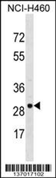
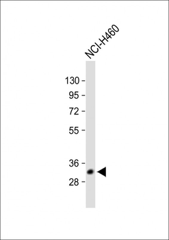
VDAC3 Antibody (Center) western blot analysis in NCI-H460 cell line lysates (35 ug/lane). This demonstrates the VDAC3 antibody detected the VDAC3 protein (arrow).
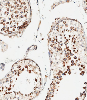
Immunohistochemical analysis of paraffin-embedded human testis tissue was performed on the Leica BOND RXm. Samples were incubated with primary antibody (1/500) for 1 hours at room temperature. A undiluted biotinylated CRF Anti-Polyvalent HRP Polymer antibody was used as the secondary antibody.
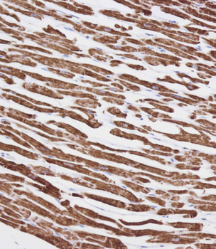
Immunohistochemical analysis on paraffin-embedded Human heart tissue. Tissue was fixed with formaldehyde at room temperature. Heat induced epitope retrieval was performed by EDTA buffer (pH9.0). Samples were incubated with primary antibody (1:100) for 1 hour at room temperature. Undiluted CRF Anti-Polyvalent HRP Polymer antibody was used as the secondary antibody.
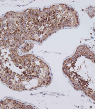
Immunohistochemical analysis on paraffin-embedded Human testis tissue. Tissue was fixed with formaldehyde at room temperature. Heat induced epitope retrieval was performed by EDTA buffer (pH9.0). Samples were incubated with primary antibody (1:100) for 1 hour at room temperature. Undiluted CRF Anti-Polyvalent HRP Polymer antibody was used as the secondary antibody.
All lanes: Anti-VDAC3 Antibody (Center) at 1:1000 dilution. Lane 1: Human heart lysate. Lane 2: NCI-H460 whole cell lysate. Lane 3: A431 whole cell lysate. Lane 4: U-2OS whole cell lysate. Lane 5: Mouse testis lysate. Lane 6: Rat brain lysate.Lysates/proteins at 20 µg per lane. Secondary Goat Anti-Rabbit IgG, (H+L), Peroxidase conjugated at 1/10000 dilution. Predicted band size: 31 kDa. Blocking/Dilution buffer: 5% NFDM/TBST.
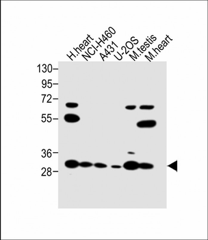
All lanes: Anti-VDAC3 Antibody (Center) at 1:1000 dilution. Lane 1: Human heart lysate. Lane 2: NCI-H460 whole cell lysate. Lane 3: A431 whole cell lysate. Lane 4: U-2OS whole cell lysate. Lane 5: Mouse testis lysate. Lane 6: Mouse heart lysate.Lysates/proteins at 20 µg per lane. Secondary Goat Anti-Rabbit IgG, (H+L), Peroxidase conjugated at 1/10000 dilution. Predicted band size: 31 kDa. Blocking/Dilution buffer: 5% NFDM/TBST.
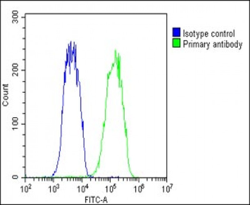
Overlay histogram showing Hela cells stained (green line). The cells were fixed with 2% paraformaldehyde (10 min) and then permeabilized with 90% methanol for 10 min. The cells were then icubated in 2% bovine serum albumin to block non-specific protein-protein interactions followed by the antibody (1:25 dilution) for 60 min at 37°C. The secondary antibody used was Goat-Anti-Rabbit IgG, DyLight 488 Conjugated Highly Cross-Adsorbed (1583138) at 1/200 dilution for 40 min at 37°C. Isotype control antibody (blue line) was rabbit IgG1 (1 μg/1x10^6 cells) used under the same conditions. Acquisition of >10, 000 events was performed.
Quick Database Links
UniProt Details
− No UniProt data available
NCBI Reference Sequences
−Associated Accession Numbers
Curated reference sequences for the gene transcript and protein product| Protein | NP_005653.3, NP_001129166.1 |
|---|
Documents Download
Datasheet
Product Information
Request a Document
Protocol Information
WB
Western Blot (IB, immunoblot)
IHC-P
Immunohistochemistry Paraffin
FC
Flow Cytometry
Li, Ying et al. Changes in the mitochondrial protein profile due to ROS eruption during ageing of elm (Ulmus pumila L.) seeds Plant Physiol Biochem, 114, 72-87 (2017)
VDAC3 Antibody (Center) (orb140381)

- 0.0
Based on 0 reviews
Participating in our Biorbyt product reviews program enables you to support fellow scientists by sharing your firsthand experience with our products.
Login to Submit a ReviewAvailable Sizes
Select a size below
Choose Conjugation or Carrier Free Version
Free Secondary Antibody (20 ul)0/0
Please add an antibody product to your cart first.
 Usually dispatched within 5-10 working days