You have no items in your shopping cart.
Featured
KO/KD
Validated
Validated
Description
Research Area
Cell Biology
Images & Validation
−Item 1 of 10
| Tested Applications | ELISA, ICC, IF, KO/KD Validated, WB |
|---|---|
| Reactivity | Human, Mouse, Rat |
Key Properties
−| Antibody Type | Primary Antibody |
|---|---|
| Host | Rabbit |
| Clonality | Polyclonal |
| Isotype | IgG |
| Immunogen | Anti-DR4 antibody (orb1239343) was raised against a peptide corresponding to 20 amino acids near the amino terminus of human DR4. The immunogen is located within the first 50 amino acids of DR4. |
| Target | TNFRSF10A |
| Molecular Weight | Predicted: 50kDObserved: 55kD (Post-modification: 1 N-linked glycosylation) |
| Purification | DR4 Antibody is affinity chromatography purified via peptide column. |
| Conjugation | Unconjugated |
Storage & Handling
−| Storage | Maintain refrigerated at 2-8°C for up to 2 weeks. For long term storage store at -20°C in small aliquots to prevent freeze-thaw cycles. |
|---|---|
| Form/Appearance | Liquid |
| Buffer/Preservatives | DR4 Antibody is supplied in PBS containing 0.02% sodium azide. |
| Concentration | 1 mg/ml |
| Expiration Date | 12 months from date of receipt. |
| Disclaimer | For research use only |
Alternative Names
−DR4 Antibody: DR4, APO2, CD261, TRAILR1, TRAILR-1, DR4, Tumor necrosis factor receptor superfamily member 10A, Death receptor 4, TRAIL receptor 1
Similar Products
−- Item 1 of 11
TNFRSF10A Antibody [orb1239340]
ELISA, ICC, IF, IHC-P, KO/KD Validated, WB
Human, Mouse, Rat
Rabbit
Polyclonal
Unconjugated
0.02 mg, 0.1 mg - Item 1 of 1
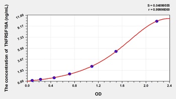
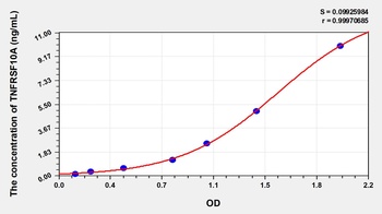
Rat Tumor Necrosis Factor Receptor Superfamily, Member 10A (TNFRSF10A) ELISA Kit [orb1146955]
Rat
0.16-10 ng/mL
0.057 ng/mL
48 T, 96 T - Item 1 of 1
Mouse Tumor Necrosis Factor Receptor Superfamily, Member 10A (TNFRSF10A) ELISA Kit [orb777287]
Mouse
0.16-10 ng/mL
0.053 ng/mL
96 T, 48 T - Item 1 of 4
TNFRSF10A Antibody [orb400932]
ELISA, IF, IHC, IP, WB
Human
Rabbit
Polyclonal
Unconjugated
100 μg, 50 μg - Item 1 of 2


Quality Guarantee
Explore bioreagents carefree to elevate your research. All our products are rigorously tested for performance. If a product does not perform as described on its datasheet, our scientific support team will provide expert troubleshooting, a prompt replacement, or a refund. For full details, please see our Terms & Conditions and Buying Guide. Contact us at [email protected].
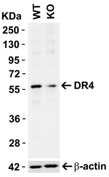
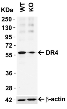
KO Validation in HeLa Cells. Loading: 10 µg of HeLa WT cell lysates or DR4 KO cell lysates. Antibodies: DR4 orb1239343 (1 µg/mL) and beta-actin orb1240312 (1 µg/mL), 1 h incubation at RT in 5% NFDM/TBST. Secondary: Goat Anti-Rabbit IgG HRP conjugate at 1:10000 dilution.
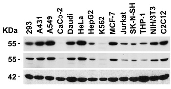
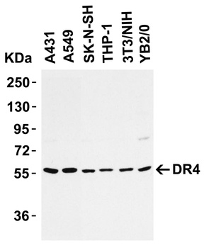
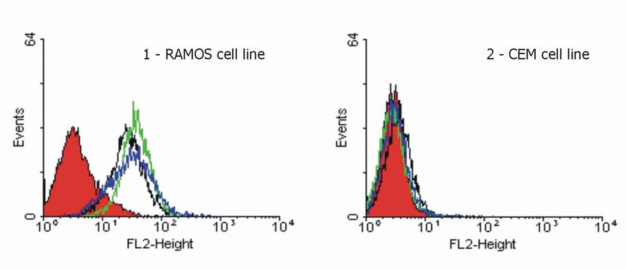
Independent Antibody Validation (IAV) via Protein Expression Profile in Cell Lines. Loading: 15 µg of lysates per lane. Antibodies: DR4 orb1239340 (1 µg/mL), DR4 orb1239343 (4 µg/mL), beta-actin (1 µg/mL), and GAPDH (0.02 µg/mL), 1h incubation at RT in 5% NFDM/TBST. Secondary: Goat anti-rabbit IgG HRP conjugate at 1:10000 dilution.
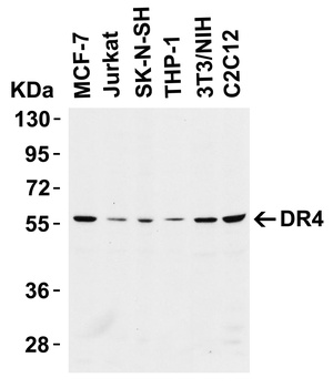
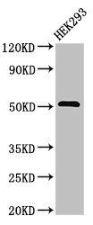
Western Blot Validation in Cell Lines. Loading: 15 µg of cell lysates per lane. Antibodies: DR4 orb1239343 (4 µg/mL), 1h incubation at RT in 5% NFDM/TBST. Secondary: Goat anti-rabbit IgG HRP conjugate at 1:10000 dilution.
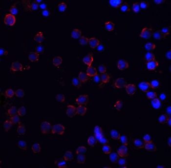
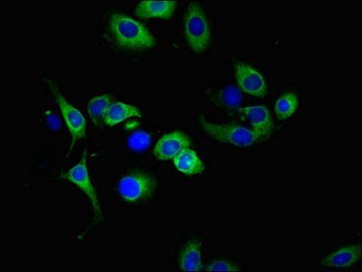
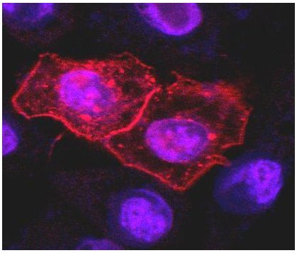
Immunofluorescence Validation of DR4 in HeLa Cells. Immunofluorescent analysis of 4% paraformaldehyde-fixed HeLa cells labeling DR4 with orb1239343 at 5 µg/mL, followed by goat anti-rabbit IgG secondary antibody at 1/500 dilution (red) and DAPI staining (blue). Image showing membrane staining on HeLa cells.
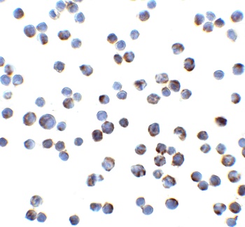
Immunocytochemistry Validation of DR4 in HeLa Cells. Immunocytochemical analysis of HeLa cells using anti-DR4 antibody (orb1239343) at 2 µg/ml. Cells was fixed with formaldehyde and blocked with 10% serum for 1 h at RT; antigen retrieval was by heat mediation with a citrate buffer (pH6). Samples were incubated with primary antibody overnight at 4°C. A goat anti-rabbit IgG H&L (HRP) at 1/250 was used as secondary. Counter stained with Hematoxylin.
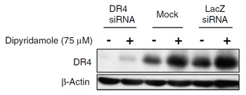
KD Validation in SW480 Cells (Goda et al., 2008). The expression of DR4 was knocked down via DR4 siRNA, 24 h latercells were treated with dipyridamole for 24 h. DR4 protein expression detected by anti-DR4 antibodies was disrupted. Dipyridamole up-regulated the expression of DR4.

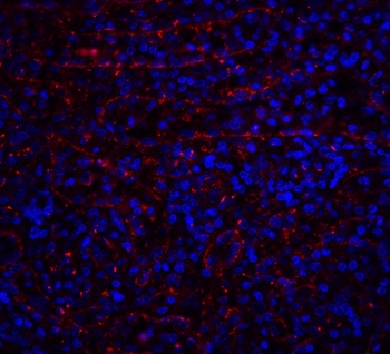
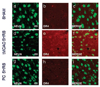
Immunofluorescence Validation of DR4 in Rat Brain (Cantarella et al., 2014). DR4 protein expression detected by anti-DR4 antibodies was increased after transient brain ischemia (tMCAO) and decreased after pre-conditioning stimulus. Confocal microscopic images displaying NeuN (a, d, g) (green), DR4 (b, e, h) (red), and Merge (c, f, i) (yellow) in the brain peri-ischemic region of rats after 5 h.
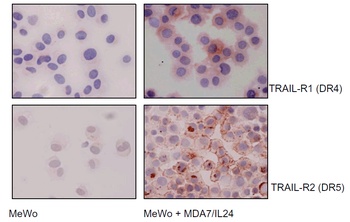
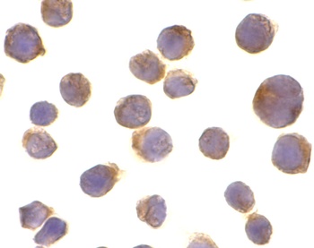
Immunocytochemistry Validation of DR4 in Human Melanoma Cells (Ekmekcioglu et al., 2008). MeWo melanoma cells were exposed to affinity-purified MDA7/IL-24. After 48 h of treatment, cells were collected and cytospins prepared for cytochemical assessment of their TRAIL receptor (R1 and R2) expression (anti-DR4 or anti-DR5, AEC, hematoxylin). Both DR4 and DR5 expression were upregulated in MeWo cells after treatment.
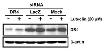
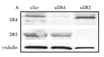
KD Validation in HeLa Cells (Horinaka et al., 2005). HeLa cells were transfected with DR4siRNA or LacZ control siRNA. At 24 h after transfection, the cells were treated with or without 20 µM luteolin for 24 h. Western blot analysis was carried out with anti-DR4 antibodies. DR4 expression was markedly reduced after DR4 knockdown.
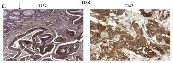
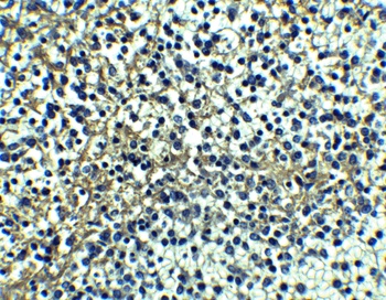
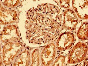
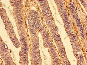
Immunohistochemistry Validation of DR4 in Human Colon Tumors (Devetzi et al., 2016). DR4 expression in human colon tumors detected by anti-DR4 antibodies. Strong immunoreactivity is shown for DR4 in T167. Moderate immunoreactivity is shown for DR4 in T187.
Documents Download
Datasheet
Product Information
Request a Document
Protocol Information
WB
Western Blot (IB, immunoblot)
IF
Immunofluorescence
ICC
Immunocytochemistry
ELISA
Enzyme-linked Immunosorbent Assay (EIA)
TNFRSF10A Antibody (orb1239343)

- 0.0
Based on 0 reviews
Participating in our Biorbyt product reviews program enables you to support fellow scientists by sharing your firsthand experience with our products.
Login to Submit a ReviewAvailable Sizes
Select a size below
Free Secondary Antibody (20 ul)0/0
Please add an antibody product to your cart first.