You have no items in your shopping cart.
Featured
Description
Research Area
Cell Biology
Images & Validation
−Item 1 of 3
| Tested Applications | ICC, IF, WB |
|---|---|
| Dilution Range | WB (1:1000), ICC/IF (1:100) |
| Reactivity | Human, Mouse, Rat |
| Application Notes |
Key Properties
−| Host | Mouse |
|---|---|
| Clonality | Monoclonal |
| Isotype | IgG2b |
| Clone No. | 6E3-2C3 |
| Immunogen | Recombinant Full length GRP170 Protein |
| Target | GRP170 |
| Molecular Weight | 170kDa |
| Purification | Protein G Purified |
| Conjugation | Unconjugated |
Storage & Handling
−| Storage | Maintain refrigerated at 2-8°C for up to 2 weeks. For long term storage store at -20°C in small aliquots to prevent freeze-thaw cycles. |
|---|---|
| Buffer/Preservatives | PBS pH 7.4, 50% glycerol, 0.1% sodium azide. Storage buffer changes when conjugated. |
| Concentration | 1 mg/ml |
| Expiration Date | 12 months from date of receipt. |
| Disclaimer | For research use only |
Alternative Names
−GRP170, HYOU1, HSP12A, HSPH4, ORP150, ORP-150, Orp150, Orp 150, Grp170, Grp 170, GRP-170, HSP 12A, Hypoxia up-regulated 1, Hypoxia up regulated 1, Hypoxia upregulated 1, hypoxia up-regulated 1 precursor, Hypoxia up-regulated protein 1, 150 kDa oxygen-regulated protein, 150kDa oxygen regulated, 170 kDa glucose-regulated protein, 170 kDa glucose regulated protein, glucose regulated protein 170, Glucose regulated 170, DKFZp686N08236, FLJ94899, FLJ97572, HYOU1_HUMAN, oxygen regulated protein (150kD)
Similar Products
−- Item 1 of 10
ORP150/HYOU1 Rabbit Polyclonal Antibody [orb865410]
ELISA, ICC, IF, IHC, WB
Human, Mouse, Rat
Rabbit
Polyclonal
Unconjugated
100 μg - Item 1 of 1
- Item 1 of 3
- Item 1 of 3
- Item 1 of 3


Quality Guarantee
Explore bioreagents carefree to elevate your research. All our products are rigorously tested for performance. If a product does not perform as described on its datasheet, our scientific support team will provide expert troubleshooting, a prompt replacement, or a refund. For full details, please see our Terms & Conditions and Buying Guide. Contact us at [email protected].
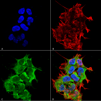
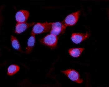
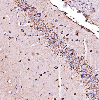
Immunocytochemistry/Immunofluorescence analysis using Mouse Anti-GRP170 Monoclonal Antibody, Clone 6E3-2C3. Tissue: Neuroblastoma cell line (SK-N-BE). Species: Human. Fixation: 4% Formaldehyde for 15 min at RT. Primary Antibody: Mouse Anti-GRP170 Monoclonal Antibody at 1:100 for 60 min at RT. Secondary Antibody: Goat Anti-Mouse ATTO 488 at 1:100 for 60 min at RT. Counterstain: Phalloidin Texas Red F-Actin stain; DAPI (blue) nuclear stain at 1:1000; 1:5000 for 60 min RT, 5 min RT. Localization: Endoplasmic Reticulum, Endoplasmic Reticulum Lumen. Magnification: 60X. (A) DAPI (blue) nuclear stain. (B) Phalloidin Texas Red F-Actin stain. (C) GRP170 Antibody. (D) Composite.

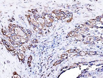
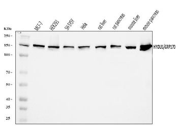
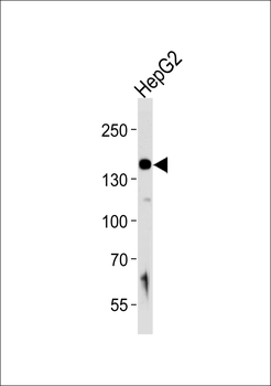
Western Blot analysis of Human Embryonic kidney epithelial cell line (HEK293) lysates showing detection of ~170 kDa GRP170 protein using Mouse Anti-GRP170 Monoclonal Antibody, Clone 6E3-2C2. Lane 1: Molecular Weight Ladder (MW). Lane 2: HEK-293 cell lysate. Load: 20 μg. Block: 2% BSA and 2% Skim Milk in 1X TBST. Primary Antibody: Mouse Anti-GRP170 Monoclonal Antibody at 1:1000 for 16 hours at 4°C. Secondary Antibody: Goat Anti-Mouse IgG: HRP at 1:100 for 60 min at RT. Color Development: ECL solution for 6 min in RT. Predicted/Observed Size: ~170 kDa. Other Band (s): 100 kDa.

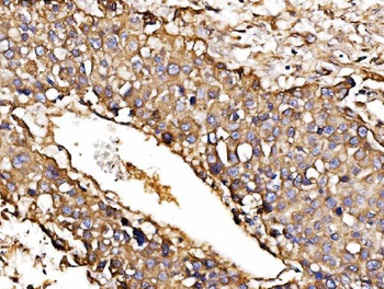
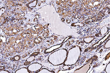
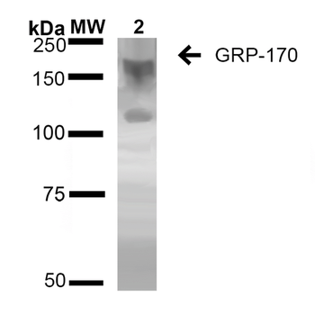
Western Blot analysis of Rat Liver showing detection of ~170 kDa GRP170 protein using Mouse Anti-GRP170 Monoclonal Antibody, Clone 6E3-2C3. Lane 1: Molecular Weight Ladder (MW). Lane 2: Rat Liver cell lysate. Load: 20 μg. Block: 2% BSA and 2% Skim Milk in 1X TBST. Primary Antibody: Mouse Anti-GRP170 Monoclonal Antibody at 1:1000 for 16 hours at 4°C. Secondary Antibody: Goat Anti-Mouse IgG: HRP at 1:100 for 60 min at RT. Color Development: ECL solution for 6 min in RT. Predicted/Observed Size: ~170 kDa.
Quick Database Links
UniProt Details
− No UniProt data available
NCBI Gene Details
− No NCBI Gene data available
NCBI Reference Sequences
−Associated Accession Numbers
Curated reference sequences for the gene transcript and protein product| Protein | NP_006389.3 |
|---|
Documents Download
Datasheet
Product Information
Request a Document
Protocol Information
WB
Western Blot (IB, immunoblot)
IF
Immunofluorescence
ICC
Immunocytochemistry
GRP170 Antibody (orb376699)

- 0.0
Based on 0 reviews
Participating in our Biorbyt product reviews program enables you to support fellow scientists by sharing your firsthand experience with our products.
Login to Submit a ReviewAvailable Sizes
Select a size below
Choose Conjugation or Carrier Free Version
Free Secondary Antibody (20 ul)0/0
Please add an antibody product to your cart first.