You have no items in your shopping cart.
Featured
Description
Research Area
Neuroscience
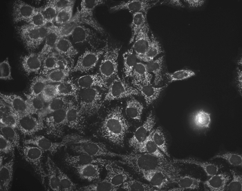
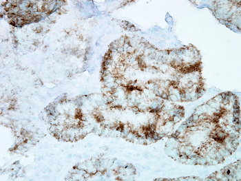
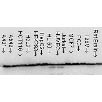
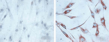
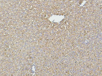
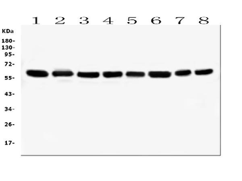
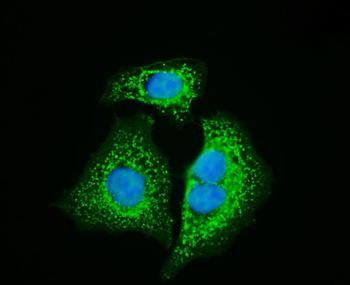
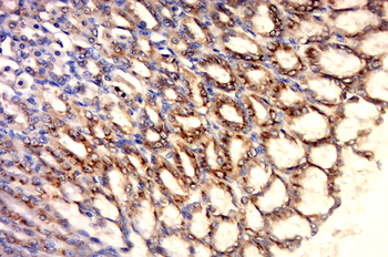
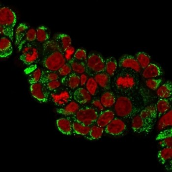
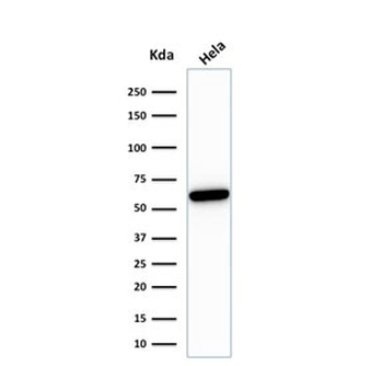
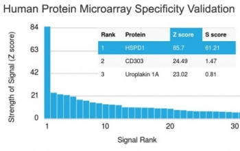
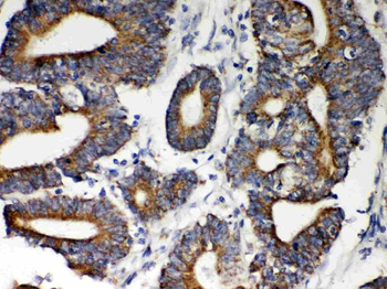
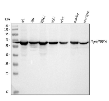
Images & Validation
−
Item 1 of 5
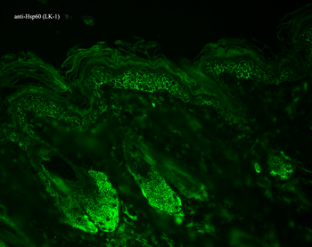
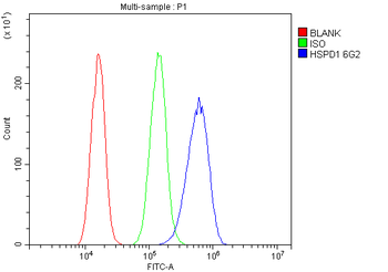
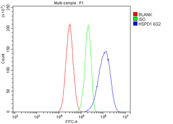
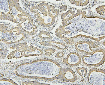
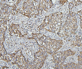
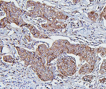
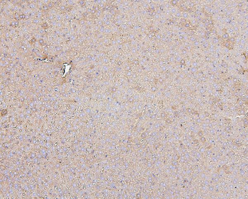
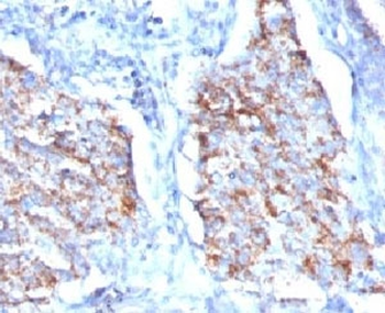
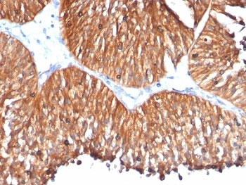
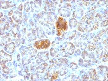
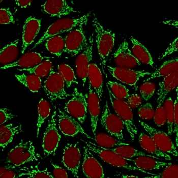
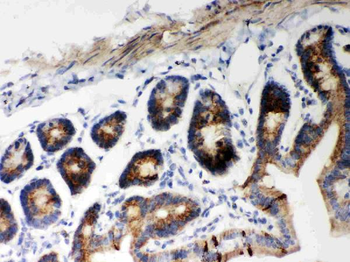
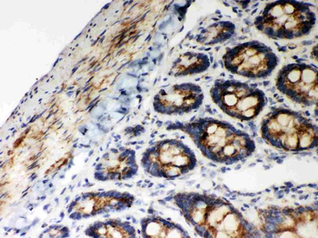
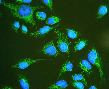
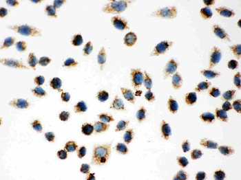
| Tested Applications | ELISA, FC, IHC, IP, WB |
|---|---|
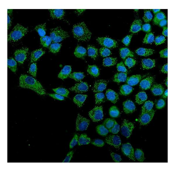
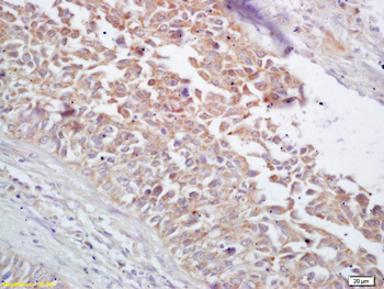
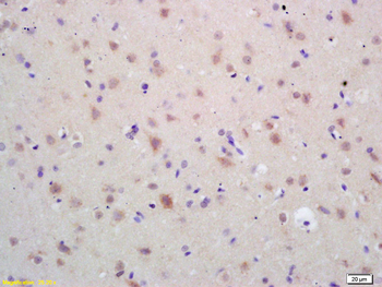
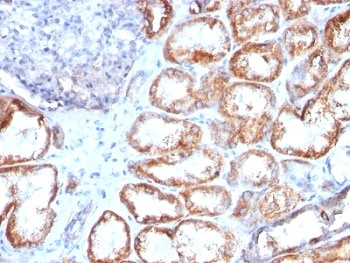
| Dilution Range | WB (1:20000), IHC (1:100), ICC/IF (1:100), IP (1:200); optimal dilutions for assays should be determined by the user. |
| Reactivity | Bovine, Canine, Drosophila, Frog, Gallus, Guinea pig, Hamster, Human, Monkey, Mouse, Other, Plant, Porcine, Rabbit, Rat, Sheep |
| Application Notes |
Key Properties
−| Host | Mouse |
|---|---|
| Clonality | Monoclonal |
| Isotype | IgG1 |
| Clone No. | LK1 |
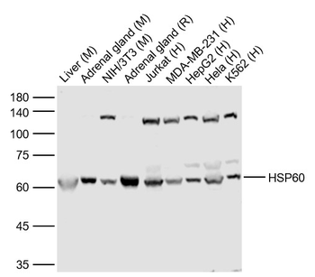
| Immunogen | Recombinant human HSP60 |
| Target | HSP60 |
| Molecular Weight | 60kDa |
| Purification | Protein G Purified |
| Conjugation | Unconjugated |
Storage & Handling
−| Storage | Maintain refrigerated at 2-8°C for up to 2 weeks. For long term storage store at -20°C in small aliquots to prevent freeze-thaw cycles. |
|---|---|
| Buffer/Preservatives | PBS, 50% glycerol, 0.09% sodium azide. Storage buffer changes when conjugated. |
| Concentration | 1 mg/ml |
| Expiration Date | 12 months from date of receipt. |
| Disclaimer | For research use only |
Alternative Names
−HSPD1, HSP60, 60 kDa heat shock protein, mitochondrial, Chaperonin 60, CPN60, HuCHA60, Heat shock protein family D member 1, GroEL homolog, mitochondrial, GROEL, HLD4, HSP 60, HSP65, SPG 13
Similar Products
−- Item 1 of 9
Hsp60/HSPD1 Mouse Monoclonal Antibody [orb570314]
FC, ICC, IF, IHC, WB
Human, Mouse, Rat
Mouse
Monoclonal
Unconjugated
100 μg - Item 1 of 5
HSP60 Rabbit Polyclonal Antibody [orb10846]
ICC, IF, IHC-Fr, IHC-P, WB
Bovine, Canine, Equine, Rabbit
Human, Mouse, Rat
Rabbit
Polyclonal
Unconjugated
50 μl, 100 μl, 200 μl - Item 1 of 8
- Item 1 of 8
- Item 1 of 6
Hsp60/HSPD1 Rabbit Polyclonal Antibody [orb251520]
ICC, IF, IHC, WB
Human, Mouse, Rat
Rabbit
Polyclonal
Unconjugated
100 μg


Quality Guarantee
Explore bioreagents carefree to elevate your research. All our products are rigorously tested for performance. If a product does not perform as described on its datasheet, our scientific support team will provide expert troubleshooting, a prompt replacement, or a refund. For full details, please see our Terms & Conditions and Buying Guide. Contact us at [email protected].
Quick Database Links
UniProt Details
− No UniProt data available
NCBI Gene Details
− No NCBI Gene data available
NCBI Reference Sequences
−Associated Accession Numbers
Curated reference sequences for the gene transcript and protein product| Protein | NP_002147.2 |
|---|
Protocol Information
WB
Western Blot (IB, immunoblot)
IHC
Immunohistochemistry
FC
Flow Cytometry
ELISA
Enzyme-linked Immunosorbent Assay (EIA)
IP
Immunoprecipitation
Available Sizes
Select a size below
Free Secondary Antibody (20 ul)0/0
Please add an antibody product to your cart first.